Protracted Withdrawal is Sensitivity to the Sulfite Paradox and Modern-day Lions

Protracted Withdrawal is Sensitivity To the Sulfite Paradox and Modern-day Lions

© 2025 by Meredith Arthur, MS, RD, LD is licensed under CC BY-NC-SA 4.0 

Last night, Jan’s husband held off washing the dishes after dinner because Jan wanted a super hot bath as a treat after a long day of caring for her rambunctious children. Jan suffers from extreme premenstrual syndrome and has been diagnosed with premenstrual dysphoric disorder (PMMD), which makes her easily irritated with her children before the onset of her period. She was prescribed selective serotonin reuptake inhibitors (SSRI) and Xanax, a benzodiazepine, to help with her anxiety and mood swings during high school and continued them through college. After graduation and getting married, she decided to go holistic in preparation for having children, so she weaned off the pharmaceuticals but experienced severe withdrawal symptoms. She and her husband, Dave, had three lovely children in the past five years after she successfully got off of benzos and recovered from withdrawal. Those scary days haunted her, and she always worried about relapsing into the nightmare. Now, she naturally treats her PMMD, including soothing baths to ease tension. Her baths were usually hot but not as hot as she wanted, and her husband knew that and knew she truly needed something to calm her very on-edge body that night. 

Jan felt a bit dizzy after the bath but headed off to bed immediately and fell fast asleep until around 2 AM when, to her horror, her heartbeat woke her up. The familiar pounding sensation couldn’t be real! How is this possible? She sat up in bed and immediately felt nauseous. She was going to vomit. She stood up and realized her feet were on fire, and her muscles were so tight that each step felt like stabbing pains. As she stumbled to the bathroom, her anxiety increased, and she felt the air hunger hit hard.  She screamed in horror, “No. It’s back. I’m not okay. I’m not okay. I’m not okay,” while she sank to the floor next to the toilet. Dave, startled from sleep, ran into the bathroom and squatted next to her. He was scared to touch her because he knew she might not be able to handle his touch now. He had spent years waiting to be able to feel her soft skin with his hands, and tears streamed down his face as he realized it would be a long time before her body felt safe enough for a hug. They both stared at each other in terror. How could this be possible? Life was so good. 

***********************************************************************************************************Often, in the protracted withdrawal community, people are told, “Only time can heal.” Well, if a moment in time like the one above can undo healing, how is time the only thing that can heal? ***********************************************************************************************************

Time does heal the brain. It takes time to restore GABA-A synapses, but why does a moment in time like the one above seemingly undo all the healing, causing setbacks sometimes to the point of being in acute withdrawal? 

One metabolite that all humans share in varying degrees that can damage GABA-A synapses when made in excess in the human body is a derivative of sulfite, s-sulfocysteine (SSC). What if the quantity of s-sulfocysteine and sulfite determines your ability to stay healed in your body at any given moment? What if these moments in time that undo all the healing that time provides is an uptick in a naturally occurring compound that we all make every day? What if finding and avoiding triggers that increase this compound and getting better at sulfite metabolism is the key to getting and staying healed?

Knowledge is the antidote to fear,- Knowledge, Use and Reason, with its higher aids. – Ralph Waldo Emerson

Meredith Arthur, MS, RD, LD believes that protracted withdrawal is an increased sensitivity to the Moco Steal and Sulfite Paradox, described by herself and Jenny Jones, PhD. This paradox is caused by an interplay between an immune and hypoxia pathway called hypoxia-inducible factor one alpha (HIF-1alpha) and a pathway called transsulfuration, where hydrogen sulfide, glutathione, taurine, thiosulfate, and sulfate are made. The triggering of the HIF-1alpha pathway leads to an increase in an enzyme called cysteine dioxygenase (CDO), leading to an overall rise in sulfite production, which must be metabolized to sulfate. Sulfate plays beneficial roles in the body, such as making healthy collagen, connective tissue, and myelin (a coating on nerves that makes them run smoothly), as well as being used for detoxifying toxins and hormone metabolism. 

Sulfite, however, is destructive to the body. To deal with excess sulfite that isn’t made into sulfate, the body will bind it to cystine (two cysteine amino acids bound together) to produce s-sulfocysteine (SSC). Everyone makes SSC daily in varying amounts. Chris Masterjohn, PhD, provides an excellent summary of the variations in SSC among humans, and says, “You would think something that is present in everyone’s urine and varies 50-fold that happens to activate NMDA receptors and cause neurological degeneration could be a powerful explanation for a major part of the variability in trait anxiety, muscle tension, ease of being startled, and the risk of neurological and psychiatric disorders.” Meredith and Jenny agree with Chris’s astute observation and believe that individuals in their Facebook support group, The MoCo Steal: Escaping From the Sulfite Paradox, tend to be more sensitive to the damaging effects of SSC, but also sulfite itself. They believe that our members who have protracted withdrawal are struggling with elevated levels of SSC and sulfite toxicity.

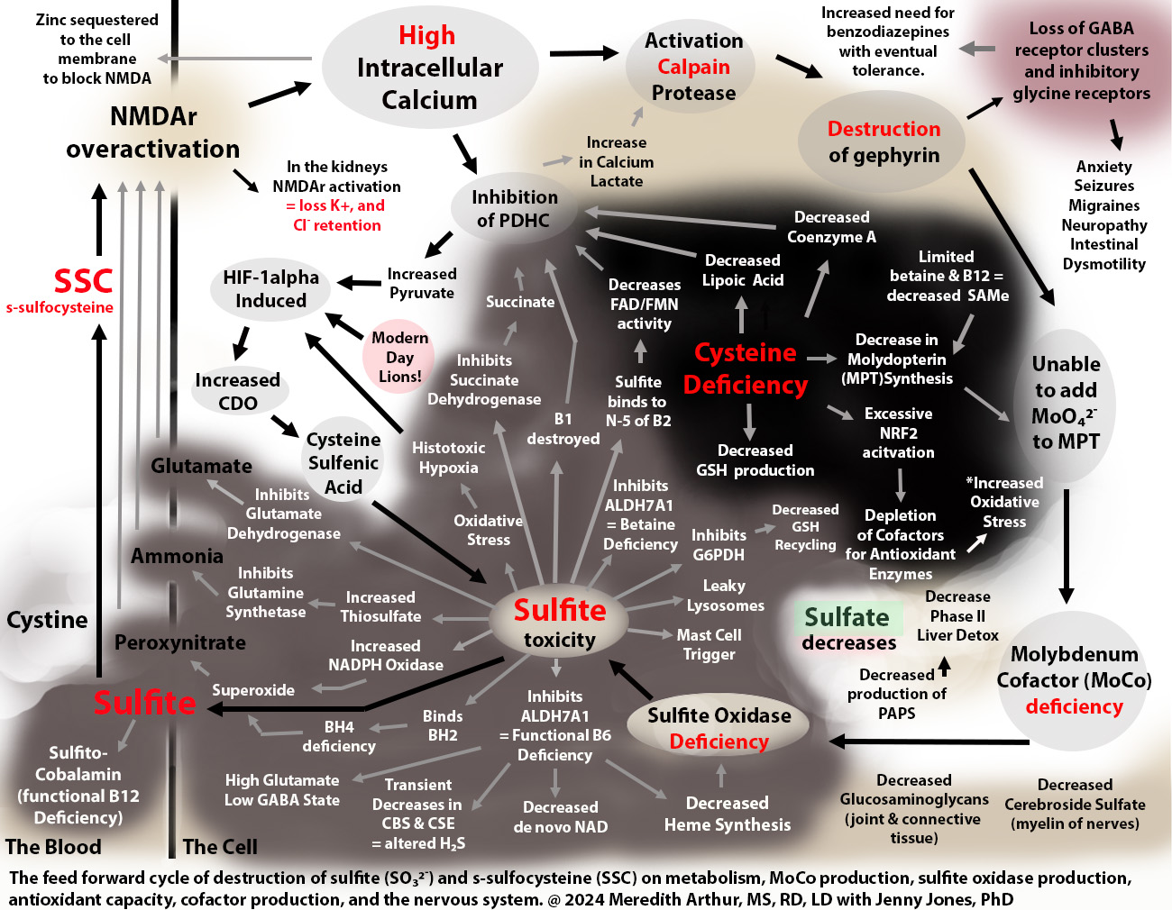
Why, then, would humans even make this damaging version of sulfite? Meredith believes that during a period of increased need for the HIF-1alpha pathway, SSC can serve as an activator of NMDA receptors found in the brain, causing mental alertness and alterations in the metabolism of other organs throughout the body that have NMDA receptors. These alterations are required during a period of metabolic or immune stress. The issue arises when HIF-1alpha remains on for too long, leading to prolonged increases in CDO and sulfite, as seen in the diagram below. 

When excess sulfite accumulates, it can inhibit glutamate dehydrogenase activity, leading to an accumulation of glutamate and nerve damage. At the same time, sulfite can cause a functional B6 deficiency that causes a loss of the conversion of glutamate to GABA. GABA calms down nerve excitation. Even worse, as shown in the upper right-hand corner of the diagram above, excess SSC leads to a loss of GABAergic synapses. SSC or other metabolites over-activate NMDA receptors in the brain, leading to an excess amount of calcium entering cells. This activates calpain protease, which breaks down gephyrin, a glue that holds the GABA-A receptors in place. Loss of gephyrin leads to a high excitatory state in the brain due to the excessive activation of NMDA receptors without an adequate inhibitory response from GABA-A synapses. This loss of the glue, gephyrin, that holds GABA-A synapses in place is a contributor to insomnia, anxiety, nerve pain, and seizure disorder

In addition, sulfite inhibits the process of methionine salvage by binding to B12 as well as by inhibiting the production of betaine, both of which are needed for restoring methionine levels to provide s-adenosyl methionine (SAM) required to make the molybdenum cofactor that is necessary for the enzyme sulfite oxidase. SSC, again, destroys gephyrin. Gephyrin isn’t just a glue that holds GABA receptors in place, but it is also the glue that sticks molybdenum into molydopterin to make MoCo. Without gephyrin, we can’t incorporate molybdenum into MoCo, no matter how much molybeneum is consumed. Thus, once out of control, sulfite prevents its metabolism by destroying the ability to make the cofactor for sulfite oxidase, MoCo, needed for its metabolism. 

SSC Contributes to Tolerance

Individuals with high SSC levels feel anxious and have muscle spasms, insomnia, and sometimes seizures. They will often be prescribed benzodiazepines, which bind to what little GABA-A receptors are left and can calm down the excitatory actions of SSC. However, at some point, due to excessive activation of NMDA receptors and loss of gephyrin, the GABA-A receptors are no longer present in large enough quantities for any benzodiazepines to have a therapeutic effect. It is at this point that Meredith thinks that individuals on benzodiazepines experience “tolerance” and are weaned off of the drug as it is no longer working. 

S-Sulfocysteine Helps Us Escape A Lion

One way to think of SSC is that it is a chemical that helps us escape a lion. Of course, none of us are escaping actual lions right now. However, modern-day lions induce the HIF-1alpha pathway, as depicted in the diagram below on the left. Any of these lions can lead to the overproduction of SSC and sulfite with the downstream damaging effects on health and metabolism shown in the diagram. 

Even stressful moments, such as the loss of a loved one, the loss of a job, a divorce, or a massive unexpected bill, can lead to increases in adrenaline and norepinephrine that encourage unhealthy gut bacteria to grow. These unhealthy bacteria lead to bacterial toxins and leaky gut that induce the HIF-1alpha pathway and CDO and sulfite production. So, even that stressful moment in life can lead to an uptick in sulfite and SSC production and anxiety.

Jan, in the introduction, experienced low blood pressure due to a very hot bath while experiencing PMMD. Right before a woman’s period, the body has a surge of estrogen, which induces the HIF-1alpha pathway, but low blood pressure also induces the HIF-1alpha pathway.  This leads to increased expression of CDO, resulting in more sulfite and SSC, leading to symptoms of protracted withdrawal. Often, individuals with protracted withdrawal are unable to pinpoint the exact cause of their regression into torment, and this may be due to HIF-1alpha being induced by many different unforeseen alterations in metabolism and sometimes circumstances beyond a person’s control. However, Dr. Jones and Meredith are working together to identify HIF-1alpha triggers, of which many are listed in the diagram above, to mitigate the uptick in this pathway in individuals vulnerable to sulfite and SSC toxicity. 

Too much SSC because HIF-1alpha is stuck in the “on” mode!

Meredith believes that HIF-1alpha is stuck in an “on” mode for too long, leading to excess SSC production, but in the past, it was beneficial when truly running from danger. For the sake of simplicity, let’s use low oxygen and running from a lion as an example. While running from a lion, oxygen is used up quickly. When oxygen goes low in a cell, it induces the HIF-1alpha pathway, which increases CDO activity. This results in more production of sulfite and more SSC. SSC then fires up NMDA receptors in the central and peripheral nervous systems, which gives a sense of alertness, allowing us to identify if any more lions are hiding in the bushes ahead of us. This HIF-1alpha pathway also increases the total amount of blood vessels in the muscles so that the next time the lion attacks, more oxygen will be available to those tissues so that if the escape ever turns into a long-distance sprint, our muscles will be ready. 

HIF-1alpha increases CDO activity, but it also changes metabolism so that a person makes energy quickly from glucose, which comes from carbohydrates (bread, cereal, rice, pasta, fruit, and vegetables). It also helps send glucose through a pathway called the pentose phosphate shunt that makes NADPH. NADPH helps recycle glutathione, a master antioxidant, from its used-up state (oxidized) to its fresh state (reduced). This reduced glutathione can mop up the oxidative stress that is happening. This change in metabolism can happen in any cell, including immune cells, to give them energy to fight infections. Unfortunately, excess sulfite can inhibit the enzyme that starts the pentose phosphate shunt, glucose-6-phosphate dehydrogenase, causing low levels of NADPH. Sulfite can also bind to oxidized glutathione, making recycling it back to reduced glutathione impossible. 

Excess sulfite, however, should be metabolized to sulfate, not remain as sulfite. If done well, the intersection of HIF-1alpha and the transsulfuration pathway helps to increase the total amount of sulfate available in the body. This is if the enzyme that helps to make sulfate, sulfate oxidase (SUOX), is working well. SUOX is the end-game enzyme needed to prevent a feed-forward cycle that keeps the HIF-1alpha pathway turned on. This is because both SSC and sulfite will induce the HIF-1alpha pathway, which is explained in more detail in the summary of the MoCo Steal Leads to a Sulfite Paradox. Unfortunately, someone with acquired or genetic SUOX deficiency doesn’t make sulfate in this pathway. Instead, they have a build-up of sulfite, thiosulfate, and SSC.

How Benzodiazepines Lead to Overactivation of HIF-1alpha and a Moco Steal

MoCo is a cofactor made with Molybdenum (Mo), a mineral that is used only for five enzymes in the human body: xanthine oxidase (XO), xanthine dehydrogenase (XDH), aldehyde oxidase (AOX), sulfite oxidase (SUOX), and mitochondrial amidoxime reducing compound (mARC). All of these enzymes are necessary for metabolism, but SUOX is crucial to prevent sulfite toxicity. Please note that SUOX is the only enzyme that also requires heme for production. Heme synthesis is compromised in sulfite toxicity due to sulfite causing a functional B6 deficiency. This leads to the loss of pyridoxal-5-phosphate (P5P) needed for the first step in the heme synthesis pathway. Sulfite toxicity also causes the loss of lipoic acid, zinc, and copper needed for further steps in the heme pathway. 

Jenny Jones, PhD, has developed a theory on how she suffered from and acquired sulfite oxidase deficiency, resulting in sulfite toxicity and excessive levels of SSC, glutamate, and peroxynitrate, which lead to adult onset seizure disorder. Her theory, The MoCol Steal, is that due to blocks in NAD production and recycling, and various other blocks at aldehyde dehydrogenase (ALDH), she had to use more of the low affinity, high-capacity enzyme for vitamin A metabolism, aldehyde oxidase (AOX) as well as XO and XDH  that also participate in vitamin A metabolism during a low NAD state, the cofactor needed for alcohol dehydrogenase and ALDH. This caused a stealing of molybdenum cofactor towards the XOR family (shown on the left side of the diagram below) and away from sulfite oxidase (SO or SUOX, on the right side of the diagram below), leading to a loss of sulfite oxidase activity and her downward health spiral, from which she has now recovered.

Benzodiazepines can lead to Dr. Jones’s MoCo steal through alteration of immune function, resulting in an increased need for XO activity. In the gut, there are immune cells found in the gut-associated lymphoid tissue, including macrophages. Macrophages are like the beat cops of the immune system. They look out for pathogenic bacteria and viruses. Benzodiazepines alter macrophages by interacting with alpha-1 subunits on GABA-A receptors, leading to macrophages having acidic cytoplasms. This results in a decreased ability of these white blood cells to engulf and kill bacteria as well as to produce cytokines to attract other immune cells to the fight. Benzos can increase the risk for bacterial superinfections due to these alterations in macrophage activity. Benzos have also been shown to increase haemophilus parainfluenzae, a gram-negative bacteria that contains LPS and also produces H2S, which is metabolized to sulfite in the body and increases the need for molybdenum cofactor. 

The immunosuppressive effects of benzodiazepines have been of great concern in the world of critical care as these are often used for sedation in intensive care units and have led to serious infections. Even so, while macrophages are incapacitated by benzos, other immune cells, called dendritic cells, are still available to recognize pathogens through toll-like receptors. This activates a pathway called NF-KB, which turns on an enzyme that makes hydrogen peroxide as a way to fight bacteria. This enzyme, xanthine oxidase, requires MoCo for activity, which leads to Dr. Jones’s Moco Steal. Dendritic cells also produce two more cytokines, IL-1 and TNF-1alph, which also increase xanthine oxidase levels. As the bacteria damage intestinal tissues, the infected gut tissue becomes hypoxic. Hypoxia induces the HIF-1apha pathway, leading to increased CDO activity and more sulfite, but the excess need for MoCo for xanthine oxidase has shifted molybdenum away from sulfite oxidase. Thus, benzodiazepines can lead to altered immune function that leads to Jenny Jones’s MoCo Steal, explained in the diagram above.

SSRI Alter Gut Microbiota. 

Many individuals struggling with modern-day lions get symptom relief from benzodiazepines but often are prescribed serotonin reuptake inhibitors as well for symptoms of depression. There is an ongoing debate whether the serotonin deficiency theory of depression is valid, but the fact that SSRI can speed up or slow down intestinal movements has been confirmed. At minimum, we know that altering GI transit can alter the microbiome and SSRI have been found to alter the gut microbiota. In addition, similar to benzodiazepines, at some point individuals reach a tolerance level with SSRI. Weaning off of them, however, comes with withdrawal type symptoms and ongoing nuerologocial sequalae. 

If Protracted Withdrawal is a Hypersensitivity to SSC and Sulfite toxicity, What Can I do?

The first step for dealing with an uptick in sulfite and SSC is to utilize what Jenny and Meredith call “mopper uppers” and modulators of NMDA receptors. Mopper uppers are nutrients that are capable of binding to sulfite in the body. Because they bind to sulfite, these mopper uppers are usually deficient in the body due to being bound to sulfite.  Meredith especially recommends for periods of times that she calls, “I’m not okay, I’m not okay,” that you consider this regimen with your healthcare provider to immediately mop up sulfite. If you also have insomnia, which is attributed to the overactivation of NMDA receptors, she recommends this helpful sheet to you and your healthcare provider on how to slow down NMDA receptor activity. Finally, you can be aware of the “lions” found in the diagram early in this document as well as join the MoCo Steal: Recovering From Sulfite Toxicity Support Group to share what your “lions” are and contribute to the ongoing research on how to promote the healing process. Jenny and Meredith believe that identifying and avoiding lions is a key component to healing. 

Do We Ever Fully Recover From the Sulfite Paradox/Protracted Withdrawal?

Meredith and Jenny are hopeful that now that the problem has been properly identified, we all can come to a healed state and that time is not the only healer. Unfortunately, the crossroads of HIF-1alpha and transsulfuration is never going away, but navigating the intersection will become easier, and preventing collisions, what those with benzo protracted withdrawal call “waves”, is possible. It’s not just time that heals, but it is time to take the fear out of protracted withdrawal. Find the lions and cage them. Mop up the sulfite. Restore lost nutrients. Slow the NMDA receptors down. Heal. 

**********************************************************************************************************

****I’m a dietitian, but probably not your dietitian. Please consult your healthcare provider before making changes to your diet, supplements, medications, or lifestyle. This is written for informational purposes and is not meant to diagnose or treat a condition. – Meredith Arthur, MS, RD, LD*****

Interpretation of Organic Acid Testing In The Setting of Suspected Sulfite Toxicity

Alterations in organic acid testing results can be interpreted in many ways. Here is a sulfite focused interpretation of changes in organic acid testing.

*****THIS IS NOT MEDICAL ADVICE – PLEASE CONSULT WITH YOUR PROVIDER BEFORE MAKING ANY CHANGES*****

Marker#59 2-hydroxybutyric acid is typically thought to be from increased CBS activity to produce more glutathione. However, this could be the product of a block at BKCDH as the alpha-ketobutyrate is metabolized to propionyl CoA via the enzyme branched chain keto acid dehydrogenase (needs B1, CoA, lipoate, FAD, NAD – of which sulfite toxicity destroys all of these).  Alpha ketobutyrate will then be forced to produce 2-hydroxybutyric acid. In sulfite toxicity due to SUOX deficiency, it has been shown that CBS activity is negligible. Most of the H2S is coming from 3-MST. https://pmc.ncbi.nlm.nih.gov/articles/PMC7711302/pdf/main.pdf   Another possibility is a competition between BCKA and alpha ketobutyrurate for BCKDH due to the catabolism of BCAA to restore TCA cycle metabolism through succinyl CoA repletion. This has been shown to occur in this study. In theory, this would lead to increased propionyl CoA levels, competition for PCC, and possibly a build-up of butyryl CoA (see Marker #45 below). 

Marker#24 Succinate could be FAD deficiency, itaconate (but will have high #30 3-methylglutaric acid as well if itaconate), but could also be inhibition of succinate dehydrogenase by free sulfite. Normal succinate doesn’t mean you are not having a sulfite issue. It could be that you are deficient in B12 (mopping up sulfite), preventing the flux of methylmalonyl CoA to succinyl CoA.

Marker #50 Methymalonic Acid. This marker is associated with vitamin B12 deficiency. However, if you have a deficiency in a cofactor at BCKDH (B1, CoA, lipoate, FAD, NAD), then you won’t have enough methylmalonyl CoA to show up B12 deficient. In addition, you could have competition for MUT by ethylmalonyl CoA if ACADS is inhibited by CoASSH (from glutathione deficiency, which  causes CoASH to be used for the cofactor for SQR)

Marker #45 Ethylmalonic Acid. This could be high due to excess H2S, a known inhibitor of ACAD (SCAD), but we should not have excess butyrate in our general circulation. This may indicate a leaky gut. It can also mean that ACADS is being inhibited by CoASSH because of SEVERE GLUTATHIONE deficiency.  In addition, this marker could be elevated due to increased BCAA catabolism, causing a tying up of propionyl-CoA carboxylase and making it less available for butyryl-CoA metabolism. 

Marker #47, 48, 49  Adipic, Sebacic, Suberic acid. These are signs of MCAD. This could be because HIF-1alpha is currently active, either due to an immune response or hypoxia. HIF-1alpha inhibits MCAD activity. Sebacic acid is a great antimicrobial. Suberic acid is great at counteracting LPS toxins.  So….It’s a good-ish thing? Not sure about adipic acid. Needs more research.

Modern Day Lions That Induce HIF-1alpha Leading to Increase Burden on the End Game Enzyme, Sulfite Oxidase, and its Cofactor, MoCo.

We all come to the Crossroads of HIF-1alpha and the transsulfuration pathway every single day. Will your flight from the lion end in sulfate or sulfite?

S-Sulfocysteine Helps Us Escape the Lion

One way to think of S-sulfocysteine (SSC), the combination of sulfite and cystine in the blood, is that it is a chemical that helps us escape a lion. Of course, none of us are escaping actual lions right now. However, we do have modern-day lions that induce the HIF-1alpha pathway which are listed to the right.

Even stressful moments in time, such as loss of a loved one, loss of a job, a divorce, or a massive unexpected bill, can lead to increases in adrenaline and norepinephrine that encourage unhealthy gut bacteria to grow. These unhealth bacteria lead to bacterial toxins and leaky gut that induce the HIF-1alpha pathway and CDO with resulting production of sulfite. So even that stressful moment in life can lead to an uptick in sulfite production. We then make SSC to fight the threat, the lion called, STRESS. 

Some of the “lions” listed above aren’t related to hypoxia, but the hypoxia pathway is induced by circumstances outside of low oxygen. For the sake of simplicity, let’s use low oxygen and running from a lion as an example. While running from a lion, oxygen is used up quickly. When oxygen goes low in a cell it induces the HIF-1alpha pathway which increases CDO activity. This results in more production of sulfite and more SSC. SSC then fires up NMDA receptors in the central and peripheral nervous system which gives a sense of alertness, allowing us to perceive if any more lions are hiding in the bushes ahead of us. This HIF-1alpha pathway also increases the total amount of blood vessels in the muscles so that the next time the lion attacks, more oxygen will be available to those tissues so that if the escape ever turns into a long-distance sprint, our muscles will be ready. 

HIF-1alpha increases CDO activity but it also changes metabolism so that a person makes energy quickly from glucose which comes from carbohydrates (bread, cereal, rice, pasta, fruit, and vegetables). It also helps send glucose through a pathway called the pentose phosphate shunt that makes NADPH. NADPH helps recycle glutathione, a master antioxidant, from its used-up state (oxidized) to its fresh state (reduced). This reduced glutathione can mop up oxidative stress that is happening. This change in metabolism can happen in any cell, including immune cells to give them energy to fight infections. Unfortunately, excess sulfite can inhibit the enzyme that starts the pentose phosphate shunt, glucose-6-phosphate dehydrogenase causing low levels of NADPH. Sulfite can also bind to oxidized glutathione and make recycling of it impossible. Excess sulfite, however, should be metabolized to sulfate, by the end-game enzyme, sulfite oxidase.  

The HIF-1alpha pathway’s increase of CDO may lead to more taurine if adequate vitamin B6 is available, but keep in mind that sulfite toxicity can cause a functional B6 deficiency. However, in sulfite toxicity, taurine may be produced from the breakdown of Coenzyme A. Taurine levels are typically high during sulfite toxicity. Taurine acts as a natural antioxidant in the body but is problematic for people suffering from sulfite toxicity. Taurine can help mop up oxidative stress in the synapses in the brain, which is good, but can lead to increased neurotransmitter activity, which can be problematic in people with high SSC levels. Individuals with too much SSC have overactivation of NMDA receptors. Taurine can help with oxidative stress in the brain but may cause a rebound excitatory response after taking taurine. In genetic SUOX/MoCo deficiency, high taurine levels do not appear to be able to prevent neurological damage.
As mentioned above, if done well, the intersection of HIF-1alpha and the transsulfuration pathway helps to increase the total amount of sulfate available. This is if the enzyme that helps to make sulfate, sulfate oxidase (SUOX) is working well. SUOX is the end-game enzyme needed to prevent a feed-forward cycle that keeps the HIF-1alpha pathway turned on as both SSC and sulfite will induce the HIF-1alpha pathway which is explained in more detail in the summary of the MoCo Steal Leads to a Sulfite Paradox. Unfortunately, someone with acquired or genetic SUOX deficiency doesn’t make sulfate in this pathway. Instead, they have a build-up of sulfite, thiosulfate, and SSC. The damaging effects of these compounds are summarized surrounding the silhouette of the human above.

I’m a dietitian, not a physician. Please consult with your provider before making any changes to your diet, supplements, medications or lifestyle. -Meredith Arthur, MS, RD, LD

Sulfite Mopper Uppers

Sulfite can form adducts with many enzymes and nutrients in our bodies, causing massive dysfunction. Mopping up sulfite helps to reduce the feed forward activation of HIF-1alpha that leads to more expression and activity of cysteine dioxygenase, which leads to more sulfite production, and when sulfite oxidase is compromised by decreased molybdenum cofactor production or decreased heme production needed for this enzyme, this leads to the ongoing cycle of destruction. Here is a list of possible “safe” ways to mop up sulfite or work around a major block that sulfite is causing in metabolism while trying to restore sulfite oxidase activity.

Known Sulfite Mopper Uppers

(Recommendations are general guidelines and not meant to substitute for an evaluation with a health care provider regarding your individual needs. Please consult with your provider before making changes to your diet, supplements, medications, or lifestyle.)

SubstanceEffectsSupplement?
Vitamin B12Functional B12 deficiency. Sulfite binds to the cobalt center of B12. This makes binding of methyl groups and adenosyl groups impossible. There is some concern that this can occur in the digestive tract and could be a significant source of B12 deficiency. Yes. B12 is needed for methionine salvage that helps to regenerate SAMe. SAMe is needed for making molybdenum cofactor (MoCo). 
Caution for individuals with sleep apnea, B12 is sequestered to the brain. Long-term intermittent hypoxia has been shown to cause cobalt toxicity of the brain. B12 can also induce the HIF-1alpha pathway which should be good, but in the presence of SUOX/MoCo deficiency this can lead to increased intestinal sulfite. Titrating up slowly may be needed. 
Dose:

Injections- 200 mcg hydroxocobalamin or per practitioner 

Oral – 50 mcg drop (1 drop of pure encapsulations hydrox/cobalamin) every hour. 

Oral – 250 mcg sublingual every 4 hours (avoid exposure to light as light can degrade B12 into cobalt)
RiboflavinSulfite can irreversibly bind to the ketone group of riboflavin. It has been shown to bind to flavin proteins at the N5 position on the isoalloxine ring, altering enzyme activity. It can decrease the enzyme activity of choline oxidase, glycolate oxidase, sarcosine oxidase, and d-amino oxidase.Yes.
Riboflavin can increase butyrate production in the intestines. The only caution is for those with elevated ethylmalonic acid (EMA) as butyrate is metabolized to ethylmalonyl CoA. 

Dose: Per individual tolerance. In migraines, dosing is 400 mg per day. However, spacing this dose out would be helpful to buffer sulfite better as riboflavin is lost quickly in urine. 


AVOID taking riboflavin with molybdenum as in acidic pH of the stomach, riboflavin and molybdenum can make a complex. https://digitalcommons.usu.edu/etd/7173/

****Riboflavin has been shown to form chelates with other metals such as iron, copper, nickel, cobalt and zinc. Avoiding high doses with meals would be helpful if you struggle with iron, copper, or zinc.
Betaine aldehydeBetaine aldehyde binds sulfite. During free sulfite toxicity, the enzyme choline oxidase can be inhibited by sulfite. This leads to low betaine aldehyde levels. 
Sulfite can also inhibit the enzyme ALDH7A1 leading to low production of betaine.
Not directly. There are no betaine aldehyde supplements.

Choline intake is important due to choline oxidase can make betaine aldehyde, and overall, the choline cycle is impaired by sulfite.

Dose:
1-2 eggs per day (Low heat cooking to avoid increase methionine and cysteine conversion to H2S. Avoid taking molybdenum supplement with eggs if you suspect H2S producing SIBO.)
500 mg choline supplement of choice
 
Betaine supplementation can help restore betaine needed for the methionine salvage pathway. This makes SAMe, which is required in order to make choline in the body from ethanolamine. 

Dose:
You could try eating more betaine rich foods if you do not struggle with oxalates or gluten (wheat bran and beets)

Start with 250 mg betaine anhydrous and sip (you may start lower if you are a sensitive person). You may work up to tolerating about 500 mg per day—caution is needed in individuals with slow CBS enzymes and high methionine on plasma amino acid.
Sci-Hub | Progressive cerebral edema associated with high methionine levels and betaine therapy in a patient with cystathionine β-synthase (CBS) deficiency. American Journal of Medical Genetics, 108(1), 57–63 | 10.1002/ajmg.10186
Vitamin B6Sulfite can form adducts with the aldehyde group on pyridoxal phosphate, but also sulfite inhibits ALDH7A1 involved in lysine metabolism, which leads to a build up of P6C and binding of P6C to pyridoxal phosphate making it inactive. Yes.
Avoid pyridoxine forms of vitamin B6 as these can lead to competition for cofactor sites and B6 toxicity symptoms.

B6 restores CBS and CSE activity. It can lead to a sudden surge in cysteine production through CBS as well as H2S production.
It may lead to sudden increases in polyamine synthesis. In theory, individuals with SUOX/MoCo deficiency grow bacteria to make H2S and polyamines to compensate for lack of production of these in their bodies. Increasing B6 quickly may lead to sudden H2S toxicity and polyamine toxicity. 

Dose:
Start with 5 mg P5P once/day with food.
Increase to 5 mg P5P every 4 hours with foods. 
Dihydro-biopterin(qBH2)Sulfite reacts with dihydropterin. This may be the cause of high urinary losses of biopterin and contribute to BH4 deficiency. BH4 is needed for healthy nitric oxide production as well as the conversion of phenylanine and tyrosine metabolism. Unsure.
There is not a good supplemental form of BH4 on the market. There are drugs to treat BH4 deficiency such as saropterin dihydrochloride. 

Proposed Alterations in BH4 Metabolism from Sulfite Oxidase/MoCo Deficiency

Zoey’s metabolome showed increased urinary losses of biopterin. I was asked by the research scientist if I was supplementing with biopterin at the time of submitting urine samples. I was not. I suspect the cause of her losses of urinary biopterin are due to the damaging effects of sulfite toxicity on qBH2. Sulfite can bind to quinone dihydrobiopterin causing decreased recycling of qBH2 to BH4.

Adapated from: https://pmc.ncbi.nlm.nih.gov/articles/PMC8573752/figure/F3/
Sulfite-qBH2 interaction: https://sci-hub.st/https://onlinelibrary.wiley.com/doi/10.1007/s10545-011-9279-7
Prolonged sulfite exposure effects on NADPH https://pubmed.ncbi.nlm.nih.gov/25777939/

Resolution of Hyperoxaluria Due to Sulfite Toxicity?! Good thing?

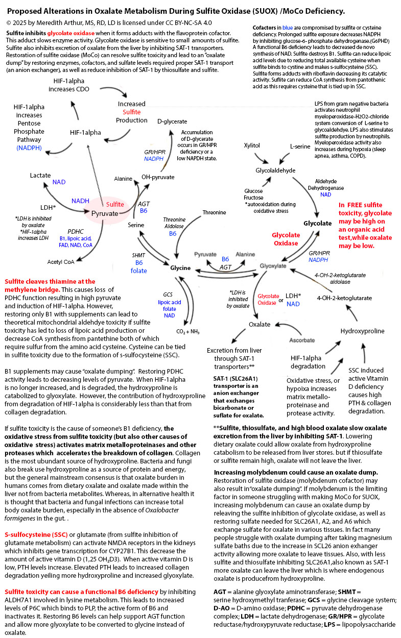
Zoey’s suddenly “cured” of hyperoxaluria as well as hyperuricemia. Is this a good thing? I think not. She has elevated glycolate levels, but low oxalate levels on her organic acid test, and her 24-hour urine oxalate levels are below the standard for someone her age as well as her urinary sulfate levels. I suspect worsening sulfite toxicity as the cause of her decreased glycolate to oxidase conversion. This isn’t a sign of improvement. It’s a sign of severe dysfunction. See the diagram below. Glycolate oxidase is inhibited by sulfite.

Zoey struggles with sleep apnea. Despite our best efforts, she rarely keeps her mask on for more than 2-3 hours per night. This means that during the night, her body increases gene transcription for HIF-1alpha in response to hypoxia. In a hypoxia state, HIF-1alpha goes to the nucleus and alters metabolism, including increasing the enzyme cysteine dioxygenase. This leads to a downstream increase in sulfite production which puts a burden on sulfite oxidase, the enzyme that metabolizes sulfite to sulfate. In addition, sleep apnea increases the activity of the enzyme xanthine oxidase. This used to be the cause of Zoey’s high uric acid levels. Xanthine oxidase uses molybdenum cofactor. Overall, sleep apnea sets the stage for Jenny Jones, PhD’s “Moco Steal” by increasing the need for more molybdenum for xanthine oxidase as well as increasing the total amount of sulfite produced by increasing CDO activity.

When the MoCo Steal from sleep apnea became extreme to the point that there wasn’t adequate molybdenum cofactor for sulfite oxidase, the sudden surge in sulfite caused a functional B6 deficiency through inhibition of ALDH7A1 resulting in a build-up of P6C. This led to decreased activity of cystathionine-beta synthase and cystathionine-gamma lyase. These two enzymes provide a significant amount of hydrogen sulfide that turns off the HIF-1alpha gene transcription. As hydrogen sulfide production decreased in Zoey’s enterocytes, she developed small intestinal bacteria overgrowth to compensate for the decrease in H2S (Greg Nigh has a theory that we grow sulfur metabolizing bacteria to provide our bodies what we need).

With the excess hydrogen sulfide in her gut, and a very oxalate restricted diet due to a history of hyperoxaluria, Zoey’s total molybdenum absorption decreased significantly due to sulfur can bind to molybdenum and when complexed with dietary copper, form a molybdenum-copper-sulfate complex that is unabsorbable. This perpetuated the problem. Over a year, her body became severely molybdenum cofactor deficient to the point that sulfite in her liver inhibited glycolate oxidase resulting in markedly high glycolate levels on her organic acids, but within range values of oxalate and below level oxalate on her 24-hour urine test.

Above you can see that Zoey’s 24-hour calcium levels have come down. She actually has decreased hypercalcemia because overall, she has improved from a vitamin A standpoint. Her serum vitamin A has dropped to 49 ug/dl which decreases osteoclasts being aggravated and breaking down bone.

Her 24 hour citrate levels have plummeted further. This is because sulfite can bind to pyruvate and prevent pyruvate metabolism to acetyl CoA. In addition, sulfite can damage all of the cofactors needed for the pyruvate dehydrogenase complex (B1, FAD, NAD, lipoic acid, and CoA). In addition, if sulfite is bound to pyruvate, it can’t be made into oxaloacetate. This leads to decreased overall levels of citrate.

Zoey’s urinary pH is quite neutral, but this is of concern to me because her blood chemistry panels over the past two years have shown hyperchloremic normal anion gap acidosis. This is consistent with renal tubular acidosis OR the actions of glutamate/s-sulfocysteine on NMDA in the kidney and activation of the sodium exchanger (ENaC). I have seen this hyperchloremic normal anion gap acidosis in many people who have suspected sulfite oxidase/moco deficiency. Activation of the ENaC results in uptake of sodium and chloride and wasting of potassium (sodium and chloride in urine below the reference range). Zoey’s urinary potassium is quite low, but her blood potassium is also low on blood draws. She has become potassium deficient and so her body is attempting to retain as much potassium as possible. During this 24-hour urine test she was getting 1000 mg of potassium citrate and 350 mg of magnesium citrate.

Zoey’s low phosphorous level is of a concern due to it may indicate intestinal malabsorption. She has a high risk for Crohn’s disease due to MBD5 deletion can increase the mRNA expression of FOLH1, that is higher in individuals with Crohn’s. The drop in her urinary sulfate below normal is consistent with sulfite oxidase deficiency. I suspect her levels are even lower because I give her magnesium sulfate foot baths every other day. The drop in her creatinine and urinary urea nitrogen indicated struggles with creatinine production (functional B6 deficiency and/or methylation decreased due to sulfite binding to B12, inhibiting betaine production, and oxidative stress damaging 5-methylfolate). Her low urinary urea nitrogen indicates a probable decrease in ornithine production needed for running the urea cycle as this is a byproduct of creatine production as well as can be made from proline but requires vitamin B6.

As you can see above, overall, Zoey is struggling with sulfite toxicity. I think that since birth (she had hypoxia in the womb due to placental abruption) Zoey has been stuck in the HIF-1alpha pathway to varying degrees her whole life. We are working on a therapeutic plan to deal with this constant, chronic uptick in sulfite production as well as strategic avoidance of sulfur foods at the time of taking mozyme forte.

References:

  1. Ghanem, Mahmoud. On the mechanistic roles of the protein positive charge close to the N(1)flavin locus in choline oxidase.
  2. Meier, Sebastian & Solodovnikova, Natalia & Jensen, Pernille & Wendland, Jürgen. (2012). Sulfite Action in Glycolytic Inhibition: In Vivo Real-Time Observation by Hyperpolarized 13C NMR Spectroscopy. Chembiochem : a European journal of chemical biology. 13. 2265-9. 10.1002/cbic.201200450.
  3. Oxidative stress from sulfite toxicity leads to loss of 5-methylfolate leading to burden on folate metabolism. Sulfite can bind to BH2 causing loss of BH4. Sulfite can lead to low levels of PLP. Pyridoxal 5’‐phosphate in cerebrospinal fluid; factors affecting concentration – Footitt – 2011 – Journal of Inherited Metabolic Disease – Wiley Online Library
  4. Glycolate Oxidase Is a Safe and Efficient Target for Substrate Reduction Therapy in a Mouse Model of Primary Hyperoxaluria Type I – ScienceDirect

High Uric Acid is ASSOCIATED with Vitamin A Toxicity

But what is vitamin A toxicity? Do we all have full livers? No. 30% of us might have full livers.

Some of us have vitamin A dysregulation which is caused by poor metabolism (low NAD state) and/or effluxing vitamin A back into the blood due to high intracellular calcium (SSC activation of NMDA, EMF activation of calcium channels, Magnesium deficiency causes STRA6 to prefer to stay on and efflux vA out into blood). High blood vitamin A levels aren’t good for sure, but they are also a squeaking wheel for major metabolic issues.

If you have HIGH retinol and LOW uric acid you have a molybdenum cofactor deficiency, and this could merely be inadequate molybdenum intake. If you take molybdenum, but still have low uric acid and high retinol, then you are struggling with making MoCo. Come join the Moco Steal group. 😉 We need to be making MoCo to have back up enzymes for metabolizing retinol and retinaldehyde. It requires several cofactors to be made.

Does vitamin A, in the form of retinol, trigger us to make uric acid? No. I would say that retinol is associated with high uric acid first due to enzyme competition. If XOR family enzymes are busy dealing with other substrates on the left of the diagram below (caffeine, high dose niacin, purines, aldehydes, etc), then it can’t play back up for ALDH enzymes metabolizing retinol and retinaldehyde.

We need MoCo for SUOX which metabolizes sulfite. Sulfite destroys thiamine. Sulfite toxicity will cause the pentose phosphate shunt pathway to prefer to send RP5 towards uric acid. If sulfite toxicity is very high it will stop the pentose phosphate shunt pathway all together by inhibiting the first enzyme, glucose-6-phosphate dehydrogenase. Having low uric acid could mean severe sulfite toxicity and severe MoCo deficiency. Having high uric acid could mean low B1 status.

B1 Deficiency Causing Increase Purine Synthesis. Another possibility is that the underlying issue is excessive shuttling or RP5 towards purine synthesis in the setting of thiamine deficiency (needed for TKT activity) due to sulfite toxicity. This sulfite toxicity could have been originally caused by high dose vitamin A causing a MoCo Steal. Sulfite immediately destroys B1, but the body will do it’s best to mop up sulfite (binds to B12, betaine aldehyde, cysteine). However, B1 deficiency can be caused by other factors too including excessive caffeine intake, too much sushi, Lasix therapy, poor oral intake, etc. B1 deficiency causes more RP5 resulting in more PRPP and the production of more uric acid. This pulls XOR enzymes away from retinol and retinaldehyde metabolism.

Does a ZERO vitamin A diet solve the uric acid problem? No. In fact, it could make it worse. The adaptative immune system uses vitamin A in the form of retinoic acid (see diagram). We do need physiological levels of retinoic acid to prevent XOR dominance happening where the innate immune system attacks bacteria and viruses with H2O2 made using Xanthine Oxidase instead of recruiting additional immune cells.

Consuming 25-50% of the RDA of vitamin A plus adding a lower dose of niacin (35 mg) at those meals seems to help my family.

****************************************

I’m a dietitian, not a doctor. I am probably not your dietitian. Please consult with a personal healthcare provider prior to making changes to you diet, medications, supplements, or lifestyle. This is written only to inform, and not to diagnose or treat a condition. This blog does not replace an evaluation by a medical professional. – Meredith Arthur, MS, RD, LD

Changes from the MoCo Steal and Sulfite Trap

Jenny Jones PhD and I have a hypothesis paper explaining how a MoCo Steal can lead to a Sulfite Trap involving the excess production of s-sulfocysteine (SSC), an NMDA agonist on steroids, and excess sulfite production. The MoCo Steal was a term adopted by Jenny as she liked your phrase, “NADPH steal” and felt her cascade into seizure disorder was caused by a shift of molybdenum cofactor away from SUOX towards AOX due to inadequate NAD for working ALDH enzymes as well as other blocks to this including high dose vitamin C. My daughter Zoey has MBD5 deletion which leads to increased expression of Mocosulfurase which may have caused her preferences to make more xanthine oxidase and aldehyde oxidase and less sulfite oxidase. As we continued to research, we found that the sulfite toxicity can be caused by the interaction of the immune system and hypoxia pathway, hypoxia-inducible factor 1 alpha which increases the expression of CDO to help with s-sulfenylation during oxidative stress as a mechanism by which to protect cells, but that instead of this metabolic switch ending in sulfate, individuals struggling with the MoCo Steal, end up in a Sulfite Trap.

Once someone is not working SUOX properly, there is a feed-forward mechanism because SSC’s robust effect on NMDA causes intracellular calcium overload leading to activation of calpain protease with subsequent degradation of gephyrin. Gephyrin is needed to add molybdenum to the molybdenum cofactor (MoCo).

Damaging effects of SSC due to excess calcium influx

  • loss of gephyrin and ability to incorporate molybdenum into MPT
  • loss of gephyrin that holds GABA receptors in place resulting in loss of functioning GABA synapses and subsequent imbalance of glutamate to GABA with effects downstream neurological effects
  • inhibition of PDHC and lactic acidosis
  • inhibition of glutamate aspartate transporter (GLAST) causing loss of glutamate uptake in neurons resulting in low brain energy levels
  • potassium and sodium wasting via over-excitation of NMDA in the kidneys with subsequent loss of the ability to uptake bicarbonate, increased H+ ions, and retention of chloride causing a non-anion gap hyperchloremic acidosis

Sulfites direct damaging effects include:

  • need for metabolizing by cytochrome C using oxygen leading to loss of mitochondrial oxygen and production of sulfite radicals. The oxidative stress triggers HIF-1alpha which increases CDO and leads to more sulfite due to the loss of SUOX activity
  • inhibition of ALDH7A1 with subsequent altered lysine metabolism resulting in a build-up of P6C and functional B6 deficiency
  • cleavage of the methylene bridge of thiamine causing thiamine deficiency
  • binding to B12 as sulfito-cobalamin causing a functional B12 deficiency and loss of MUT causing inadequate succinyl CoA and slowed heme synthesis
  • inhibiting glutamate dehydrogenase in neurons results in decreased alpha-ketoglutarate, diminished movement of the TCA cycle, decreased mitochondrial membrane potential, and decreased ATP synthesis.
  • inhibiting glutamate dehydrogenase in other cells leads to similar effects, but also loss of alpha-ketoglutarate to act as an antioxidant during oxidative stress
  • damage to lysosomes causing them to leak cyanide which ties up MPST in the production of thiocyanate. Thiocyanate causes loss of uptake of iodine into the thyroid gland and hypothyroidism

Functional B6 deficiency from ALDH7A1 inhibition causes:

  • inability to complete de novo NAD production via the kynurenine pathway
  • loss of CBS and CSE activity
  • inability to start heme synthesis due to ALAS requires B6 (or minimal ability due to triage flux of B6 towards important cellular needs)
  • decreased serine hydroxy methyltransferase (SHMT) activity leading to decreased glycine production from serine, but glycine supplements cause reactions due to acting as co-agonists to NMDA
  • severe cases lead to a lack of histamine production (resolution of histamine intolerance but at the same time severe neurological presentation with loss of motor coordination)
  • less severe cases, decreased ability to metabolize histamine with ALDH7A1 side of histamine metabolism leading to increased burden on HNMT
  • decreased glutamate to GABA conversion

Sulfite binds to cystine to make SSC which can cause cystine deficiency. This could be restored by SSC being taken up by cells and metabolized using glutathione, but the shift towards the hypoxia pathway causes more cysteine to be shuttled towards CDO and overall there is a glutathione deficiency. Due to oxidative stress, cysteine is also shuttled towards CDO to produce cysteine sulfenic acid to protect enzymes during oxidative stress so that when it resolves the cell can resume function.

Cysteine deficiency then causes…

  • loss of lipoic acid, CoA, and FAD and FMN (sulfite binds to the N-5 catalytic site of B2 causing flavin containing enzymes to be nonfunctional).
  • overactivation of NRF2 activation leading to keratinization
  • vitiligo due to failure to prevent apoptosis of melanocytes and due to sulfite can inhibit glycolate oxidase. Glycolate inhibits tyrosine hydroxylase needed to produce melanin.
  • (CoQ10 needs cysteine for production – prenylation- but this seems conserved due to needing large amounts of ubiquinol for degradation of HIF-1apha)

The MoCo Steal That Leads to A Sulfite Trap

Sulfite toxicity and cysteine deficiency collide at the crossroads of transsulfuration and the HIF-1alpha pathway.

I’m a dietitian, not a doctor. This isn’t medical advice. Talk with your healthcare provider before making changes to your diet, supplements, medication, or lifestyle.

For the past year or so Jenny Jones and I have been discussing her MoCo Steal Theory. Her theory is that due to blocks in NAD recycling and various other blocks at ALDH, she had to use more of the low affinity, high-capacity enzyme for vitamin A metabolism, aldehyde oxidase (AOX). This caused a stealing of molybdenum cofactor towards AOX and away from sulfite oxidase (SO or SUOX) leading to a loss of sulfite oxidase activity. Sulfite then caused the functional B6 deficiency that she and I collaborated on where the moonlighting enzyme ALDH7A1 was completely inhibited by sulfite causing a sort of late-onset pyridoxine-dependent epilepsy from a functional B6 deficiency.

.

Controlling glutamate is important through dietary interventions to reduce free glutamate if you struggle with seizures or migraines, but there is a sneaky glutamate-like compound being made in our bodies every day called s-sulfocysteine (SSC). It’s the dipeptide cystine bound to free sulfite. Chris Masterjohn talks of how we all have this in varying amounts.

.

Jenny, Andrew Baird, Michelle Harris, and I have been discussing that vitamin A “toxicity” is actually vitamin A “dysregulation” and overall isn’t a poisoning from retinoic acid because most of us aren’t even getting to the point of making retinoic acid due to the massive dysfunction caused by sulfite toxicity.

.

After many months of research, I have concluded that Jenny’s MoCo Steal may have happened to some of my clients in the past including my daughter. However, I noticed that they had gone beyond a MoCo Steal and had fallen into a Sulfite Trap because when we can’t metabolize sulfite, we make s-sulfocysteine (SSC) and have excess free sulfite causing damage to our mitochondria leading to a feedforward cycle of destruction that meets at the crossroads of transsulfuration and the immune and hypoxia response pathway, hypoxia-inducible factor-1 alpha (HIF-1alpha).

.

Regarding vitamin A metabolism. SSC over-activates NMDA receptors on many cells in the bodycausing large increases in intracellular calcium which then causes retinol to efflux out of cells back onto RPB4. This is the high serum A that we experience. It’s a cellular deficiency of vitamin A, but blood toxicity.

.

Although high liver vitamin A stores are still possible, I think that high serum vitamin A is the squeaking wheel on a very broken race car called Acquired Sulfite Oxidase Deficiency. This leads to intolerance to vitamin A-rich foods. The thesis linked to in the description section of the video will explain dry skin, collagen loss, and poor glucuronidation, but this is not covered in this preliminary video below which only explains how we get trapped in this pathway.

But the damage doesn’t stop at vitamin A intolerance.

Both sulfite in the mitochondria and SSC cause an increase in the activity of cysteine dioxygenase (CDO) through the activation of HIF-1alpha. Upregulation of CDO leads to shutdown of the cells through s-sulfenylation with cysteine sulfenic acid. This is a life-saving effort to protect enzymes from oxidative stress so that when the cell turns back on, the cell won’t have to start all over again. The bad part is, that leftover cysteine sulfenic acid is metabolized to sulfite and should be further metabolized to the beautiful, life-saving sulfate which is used for Phase II liver detox. Instead, we make more sulfite which leads to more mitochondrial damage as well as more SSC which causes more increases in CDO and more sulfite. It’s a trap.

.

SSC itself causes alterations in metabolism that cause the loss of the ability to make MoCo because it leads to loss of gephyrin needed to add molybdenum to molybdopterin and eventually loss of the ability to make the SUOX enzyme as well for some people. Cells outside of the central nervous system express NMDA receptors, which can respond to SSC, including hepatocytes, a major site of sulfite oxidase activity.

.

This pathway leads to cysteine deficiency from tying up cystine in the blood with sulfite. This eventually leads to the inability to make FAD due to FAD synthase needs the cofactor molybdopterin (MPT) which requires a sulfur group from cysteine. MPT is NOT molybdenum. It’s the step before molybdenum. Taking molybdenum is not a way to restore FAD synthesis. It’s not involved in that reaction. Just MPT, the empty shell of MoCo, for lack of better terms, is what is needed for making FAD.

.

And the damage to other cofactors and pathways is evident. Anything that requires cysteine will be decreased such as lipoic acid, coenzyme A, activation of NRF2, and production of CoQ10 (although I mostly see this pathway conserved, if not high on Genova testing, because people need CoQ10 for the HIF-1alpha pathway, but they aren’t using it in their mitochondria at SQR as the mitochondria are shut down trying to repair the damage from sulfite and I doubt that CBS and CSE are working much at all due to functional B6 deficiency from sulfite inhibiting ALDH7A1).

.

Activation of NMDA receptors in the kidneys causes potassium wasting and sodium wasting, but chloride retention, and inability to uptake bicarbonate from urine lead to non-anion gap acidosis which is often missed by doctors. It can also cause hyperaldosteronism and hypertension. I suspect that anyone requiring high dose thiamine for life with high dose potassium intake is a maker of excessive amounts of SSC. Free sulfite is probably the cause of many individuals’ thiamine deficiencies as sulfite cleaves thiamine at the methylene bridge essentially destroying it.

.

In the brain, SSC over-activates NMDA receptors on GABAergic synapses leading to excessive calcium influx into cells, loss of gephyrin needed to hold GABA receptors in place, and destruction of the synapses. This leads to a high excitatory state with low inhibition and can cause seizures as well as anxiety, pain, bowel dysmotility, tremors, and dysautonomia.

.So…I humbly ask for you to consider this hypothesis that I’m living out in real time. I am not trying to be a savior. I’m just sharing an awful experience and showing my love for my fellow humanity by putting this out there. I am currently stuck in the sulfite trap with my daughter, Zoey. I got here when I took garlic and NAC to lower my blood pressure and then added in rolled oats. It was a snowball effect leading to severe sulfite toxicity. I think I have a way of escaping, but I am tired.

Because of the underlying sympathetic surges that I have right now, negative comments will harm me physically. Please refrain from making negative comments about Jenny and my work until you have thoroughly investigated it and proven it wrong. I would like for all the great minds out there in our world to take over and help find a solution to this problem if they feel that my hypothesis is valid. I think I have a few good ideas, but I do not know everything. I need YOU…smart people. Help save my brain and everyone else’s, please.

,

If I am wrong on this HYPOTHESIS. I may end up on 20 medications to control the massive symptoms that I have. However, currently, Benadryl is helping. This is not a medication recommendation for anyone…you MUST go to your doctor or practitioner with this idea. This is a dangerous trap.

Benadryl blocks NMDA receptors nicely and is reducing all my symptoms, but I’m still not fixed because when I stop the Benadryl, I fall back into horrible symptoms such as anxiety, tachycardia, insomnia, high blood pressure (some have low blood pressure), pain, internal tremors, burning sensation all over my body, and more. Only time will tell if blocking NMDA digs me out of the trap. I have a few other ideas as well in the collaborative thesis paper that Jenny and I worked on.

If you aren’t having a hypertensive crisis, there is another way out. I had a client heal with a few small changes. She still has migraines and histamine reactions, but her neurological symptoms have improved immensely. More on that soon. 

CAUTION!!!!!!

Do not try to get out of this trap alone!!! Blocking NMDA restarts metabolism and some things might happen that aren’t pleasant. If you over block NMDA and then remove the block, you could have a SEIZURE. You must be aware of this risk. PLEASE WORK WITH YOUR PRACTITIONER.

Do not add NAC if you think you are stuck in this pathway until you get with a practitioner to find some way to block your NMDA receptor UNDER the SUPERVISION OF A HEALTHCARE PRACTITIONER but titrate it for your own needs and monitor your electrolytes and labs. Blocking NMDA receptors restarts metabolism, but we don’t have all the co-factors ready and I’m worried that fragile people need close monitoring. There is a risk that you don’t have all the cofactors for finishing the conversion from pyruvate to acetyl CoA. I think that this can lead to mitochondrial aldehyde toxicity. I sent a message to Dr Marrs about this. She plans to look at what I wrote on Monday.

I think whenever we’re using an NMDA blocker, UNDER SUPERVISION OF A HEALTHCARE PRACTITIONER we have to do a very gentle diet where we consistently eat only a small amount of carbohydrates every 2 to 2.5 hours so that we don’t go into full-blown lactic acidosis because lactic acidosis will get us stuck in the same pathway. Lactic acidosis could lead to the same increase in the CDO activity that I described in the video. So, it would be a low carbohydrate diet but not carbohydrate-free because to avoid gluconeogenesis as making glucose uses GTP. GTP is needed for making MPT and molybdenum cofactor.

Here is a link to the video. Here is a link to a HYPOTHESIS paper.

********************************