I have been longing for the tree of life that is in the book of Revelation. It has fruit each month, 12 different kinds, and the leaves are for the healing of the nation. I’m sure they are low oxalate!
I think I figured out why the 1980-ish study found that plantain had only 1 mg of oxalate, but new data is closer to 524 mg of oxalate. It is the increased use of glyphosate. Glyphosate is not just on our fruits and vegetables that aren’t organic. It is in the soil. It can stay in the soil for many years, but decreases as plants suck it up through their root systems. Then the plants metabolize the glyphosate to….oxalate!!! They aren’t naturally poisonous. They are trying to protect themselves from glyphosate. We also metabolize glyphosate that we consume to oxalate! There are some bacteria that can eat oxalate, but most people do not have this healthy oxalobacter formenges. Maybe it died off with glyphosate increases? I think so.
In my opinion, So our heavenly Father didn’t intent for us to have this poison in our foods. I do know of one lab, Unlocked Labs, who is working on turning on genes in bacteria so that they eat oxalate. That’s interesting. I sent them the hypothesis about NAD dependent vitamin A metabolism and how oxalate is inhibiting NAD recycling. We shall see how that goes.
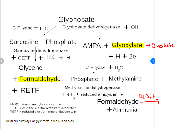
This is the metabolism of glyphosate in the human body. The body can make two products that cause damage to vitamin A metabolism. Oxalate and Formaldehyde. Oxalate impairs LDH causing decrease in NAD needed for retinol and retinal metabolism to retinoic acid. Formaldehyde puts burden on Aldehyde dehydrogenase that is needed for retinal to retinoic acid conversion.
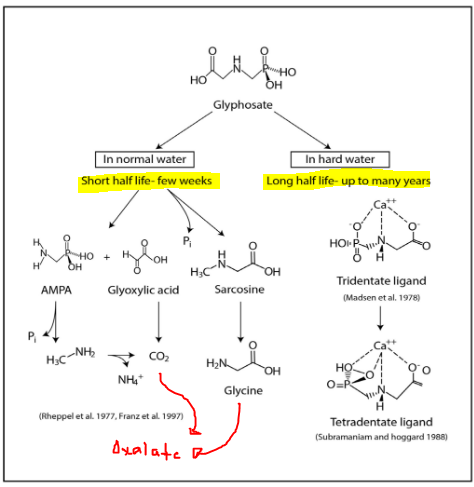
Water quality matters. This is shows that hard water (water treated with chemicals and minerals such as the water that we get from the city) will cause glyphosate to remain metabolized for many years. Normal water will result in glyphosate breaking down into metabolites that can still harm people if they are B6 or Thiamine deficienct, but it will degrade faster and not persist in the environment.
source: http://www.mdpi.com/1660-4601/11/2/2125
There’s so much more research on this persistent poison, but I think I will stop here and make some recommendations.
Avoid the dirty dozen (EWG is a good place to start)
Avoid wheat, corn, oats, and soy if not organic
Realize that the oxalate in plants is higher now likely due to the fact that glyphosate is so abundant in our soil and water supply
Forgive the dietitian who made the bad handout for not realizing this, but send her this blog.
This is a quick hypothesis on the need to correct the ability to catabolize retinoic acid (final irreversible product of vitamin A metabolism). This hypothesis is not intended to diagnose, treat, or cure a disease. It is only written for to inform. Please consult with your personal healthcare practitioner before making any changes. I am not a doctor. I am dietitian with a quirky set of skills. – Meredith Arthur, MS, RD, LD
Retinoic acid (RA) has to be catabolized and removed from the body or levels may go to high. Some symptoms that I contribute to high retinoic acid include, nausea, peeling skin, burning sensation of the skin, headaches, fatigue,
This breakdown of RA requires CYP26. There are no known inhibitors of CYP26…yet…but it does require NADPH. This can be made from NAD. In a low NAD state, you won’t have enough NADPH to be able to metabolize retinoic acid. As you work on changing habits that decrease NAD, this NADPH should improve, BUT there is a chance that you are over using all cytochrome P450 enzymes resulting in a burden on NADPH. This could lead to increased levels of RA. High RA can poison a person (think Accutane poisoning). So, I’m working on figuring out how to maximize this NADPH dependent removal of RA.
After it is metabolized by CYP26, the products of RA must go through the glucuronide pathway. There are many substrates that use this pathway. Stevia is one of them. All stevia glycosides are detoxified by this pathway. Monk fruit is also a glycoside, and so uses glucuronidation to be removed from the body. Many drugs use this pathway. So this is another possible inhibitor of properly removing retinoic acid from the body.
There is a vitamin A toxicity group on facebook that has some good information about maximizing these pathways. As I explore this more, I will add what I think is valuable information to this post. So far, these are the most important changes to make to diet (consult your healthcare provider before making changes):
increase egg yolks to four per day (Andrew Baird from the Vitamin A toxicity group recommends going slow with eggs and starting with one per day https://www.facebook.com/groups/3033243886748451/)
decrease sweeteners such as monk fruit and stevia
work on daily bowel movements, but not with Miralax. Magnesium bicarbonate may help as the bicarbonate also triggers mesothelial cells to make acetycholine independent of vagus nerve stimulation. Acetycholine is the major neurotransmitter of the gastrointestinal tract. I used to use Magnesium citrate with Zoey, but just yesterday one of my “treasures in heaven clients”, Meagan, told me she makes magnesium bicarbonate to increase magnesium levels. Well, I said, “You also just helped your GI tract move.” We learned from each other. Life is a team effort and God brings people together for the good of each other.
consider Taurine supplement to help with bile acid production
avoid glyphosate as this overloads cytochrome P450 and ties up NADPH in detoxifying it (Go Organic. Avoid non-organic wheat, soy, oats, corn. ) Check out EWG dirty dozen. https://www.ewg.org/foodnews/dirty-dozen.php
consider supplementation with calcium-D glucurate to support glucuronidation pathway (Thanks, Oskar for piping in on the Vitamin A toxicity group.)
DO NOT consider inositol to support the pentose phosphate shunt pathway that helps to make NADPH. I will make a post soon on why you should avoid inositol supplements in detail. However, to give a short explanation, inositol can impair the production of cardiolipin by posttranscriptional inhibition of enzymes in this pathway. I believe this production is already impaired by retinaldehyde accumulation that complexes with ethanolamine leading to A2E formation (lipofuscin). This decreases the availability of CDP diacylglycerol which is a precursor for cardiolipin. When cardiolipin levels decrease in the mitochondrial membrane, this can cause oxidative stress and cell death. Having high vitamin A status alone can cause this process by stealing a source of CDP diacylglycerol needed for cardiolipin production, but adding in inositol can completely inhibit this pathway. In fact, A2E accumulation in the eye actually causes damaged to cytochrome C oxidase leading to oxidative stress. Again…longer post coming soon.
Meagan recommends checking on Musclesandmotherhood on instagram for recipes and info about MgHCO3 https://www.instagram.com/_musclesandmotherhood/
This post was reviewed by Jenny Jones, PhD, human molecular genetics. I want to make sure that whatever I share with you has at least two critically thinking minds working on a problem. <3
This is not written to diagnose or treat a condition, but only for informative purposes. Please consult your doctor before stopping or starting medications or supplements, and before making dietary or lifestyle changes based on the information provided. – Meredith Arthur, MS, RD, LD
Now for the various ways that we can alter Vitamin A Metabolism…….
Oxalate
vitamin C (excess from supplements)
Glycine
Miralax or other PEG products
Thiamine deficiency
B6 deficiency
Iron deficiency
Zinc deficiency
H2 Receptor antagonists
High dose melatonin
Gut dysbiosis
Acetaldehyde (and alcohol)
Under each section there is a mechanism of action, followed by a possible solution. Of course, talk with your health care provider before making any changes.
OXALATE from plants or made from VITAMIN C or GLYCINE or Miralax (PEG) in the body can impair Vitamin A metabolism
Oxalate is a component of plants that is impossible for the body to completely break down. It is a poison. We absorb it at variable rates, but some of us make it in our bodies from vitamin C and glycine. Excess vitamin C becomes oxalate through direct breakdown and without enzymes. Usually this occurs in vitamin C over 2000 mg, but it can happen at lower doses as well. Never take vitamin C to “bowel tolerance” as this is likely actually death of the intestinal cells due to oxalate poisoning. Glycine is metabolized to oxalate in a B6 and thiamine deficient state, but when there is adequate B6 and Thiamine, it does not become oxalate.
When oxalate is high it impairs an enzyme called Lactate Dehydrogenase (LDH). We have to make some lactate to keep energy metabolism going. When the body is producing lactate, it also produces NAD+ which is what drives vitamin A (retinol and retinal) metabolism forward. What I found through a deep dive into literature is that Oxalate doesn’t directly inhibit alcohol dehydrogenase or retinol dehydrogenase or aldehyde dehydrogenase which was what I was searching for. Oxalate actually impairs lactate dehydrogenase (LDH) which lower NAD+ levels. I hypothesize that oxalate takes away the “energy” needed to drive those reactions forward by impairing LDH.
LDH is actually the last enzyme involved in the formation of oxalates. I believe that oxalate being able to have a feedback inhibition on LDH is a safety mechanism built into our human biology, but that it backfires and wreaks metabolic havoc on vitamin A metabolism and also energy metabolism.
Oxalate Pathogenic In Autism (Perhaps this is the connection! If oxalate impairs LDH, resulting in low NAD, then retinal levels increase. These complex with ethanolamine causing A2E and microglial activation resulting in neurological decline.)
Jenny Jones, PhD, pointed the article below out to me as supporting evidence for the connection between need for normal LDH reaction to restore NAD levels.
The article bleow is an excellent article! This gives the big picture of NAD production, recycling, and salvage pathways. Amazing! LDH, which is inhibited by oxalate, plays a pivotal role in NAD recycling.
NAD+ metabolism: pathophysiologic mechanisms and therapeutic potential
2. Avoid excess vitamin C in excess (variable per person, but most kids don’t need more than 500 mg per day)
3. Ensure adequate levels of B6 and thiamine (Seizure meds tend to deplete B6 – ask doctor about 50 mg of P5P, active form of B6) – ask doctor before starting supplements CAUTION – B6 in a low NAD state may be toxic.
4. Avoid glycine supplements and also collagen powders as these are high in glycine
MIRALAX can become OXALATE and also can tie up alcohol dehydrogenase and aldehyde dehydrogenase that are needed for Vitamin A metabolism
Approximately 3.7% of PEG based laxatives are absorbed. This can be metabolized by the body to glyoxylate and then to oxalate especially in a B6, Thiamine, or Niacin deficient state. This will impair LDH, subsequently lower NAD, and thus impair vitamin A metabolism, but also overall metabolism. In addition the first two steps of PEG metabolism involve alcohol dehydrogenase and aldehyde dehydrogenase. They are enzymes used in vitamin A metabolism. So Miralax may tie up these enzymes for an unknown period of time. This would be an interesting study in a rat lab.
So many people with Autism take PEG (Miralax). PEG can also cause gut dysbiosis (see below for info on bacterial steal of NAD+) Perhaps many have A2E complexes of the essential ether lipid ethanolamine due to increasing retinal levels (this is a hypothesis).
PEG with weights greater than 4000 aren’t absorbed (1960 studies), but somewhere along the way a manufacturer changed it out for PEG 3500, probably due to cost, and the researchers felt absorbing 200 ml out of 5400 ml was no big deal.
I propose that 3.7% absorbed of the PEG laxative are causing a big deal. And the unabsorbed product is causing gut dysbiosis.
Ever notice that the label says not for use in children? Also to not use more than a week?
2. Ask your doctor for alternatives such as magnesium, senna, glycerin suppositories, etc.
B6 DEFICIENCY
B6 deficiency can cause increased production of oxalate from the amino acid glycine. Also, thiamine is needed to activate B6 into pyridoxal-5-phosphate. In addition, B6 and thiamine deficiency can prevent the production of NAD from tryptophan. High losses of B6 can occur when oxalate clearing through the kidneys is high. B6 is also depleted by birth control. Many seizure medicines deplete B6.
Solution:
– Ask physician about taking P-5-P. (However, I believe at this time that supplementing P-5-P in a NAD deficient state due to alterations in dietary factors that prevent NAD recycling can cause B6 to become toxic. See this post for that hypothesis. CAUTION – B6 in a low NAD state may be toxic.
– Ask physician about thiamine supplementation
– Avoid glycine supplements or collagen powders which are high in glycine
Thiamine is needed to activate B6 to its P5P form. Thiamine is also needed to help pyruvate become lactate, which leads to adequate levels of NAD+. Thiamine deficiency can be caused by drinking too much coffee, tea, or caffeinated soda. It can also be depleted by the drugs lasix and metformin.
Solution:
– Talk with your doctor about thiamine supplementation (there are four forms, thiamine HCL, thiamine mononitrate, benfotiamine, and TTFD)
– Stop drinking so much caffeine!
– do not stop a medication without talking with your doctor
ZINC DEFICIENCY
Zinc is needed to metabolize vitamin A into retinoic acid. However, excess dietary zinc can cause a copper deficiency which can cause microcytic anemia and also neurological damage. Excess iron supplementation can cause zinc deficiency, so if you are on iron, then Zinc deficiency is possible.
Solution:
1. Ask your doctor to check ceruloplasmin and plasma zinc levels to evaluation zinc and copper
2. Ask your doctor about starting a Zinc:Copper Balance supplement. It should be about 10 to 15 mg of Zinc to every 1 mg of copper. THe amount of zinc and copper you need may need to be adjusted.
IRON DEFICIENCY
Iron deficiency impairs the mobilization of vitamin A from the liver. This can lead to a functional vitamin A deficiency, and excessive liver stores of vitamin A. The functional vitamin A deficiency worsens iron deficiency because retinoic acid, active vitamin A, down regulates the production of hepcidin by the liver, but also adipose tissues. Hepcidin increases. Hepcidin essentially locks iron into the cells of the intestine or liver so it can impair iron absorption, but also can lead to iron toxicity in the liver. (Never blindly supplement iron. Always ask for iron studies.)
Once liver capacity for vitamin A is reached, the body will increase cholesterol production in efforts to send vitamin A to fat cells for storage. If there is not enough choline to make cholesterol, fat will accumulate in the liver causing fatty liver disease. This can occur with or without iron deficiency. There are many other factors that contribute to poor vitamin A metabolism beyond iron deficiency.
Also, iron deficiency causes slowing of TCA cycle and build up of citrate which becomes a building block for triglyceride production. Less energy is also produced from food eaten because of this slowing of the TCA cycle that makes ATP in the body.
Sometimes iron deficiency that isn’t responding to iron supplementation is actually copper deficiency. If you have been on iron a long time, you should have your copper levels checked (ceruloplasmin) due to high dose iron supplements impair copper absorption. Once copper is low, then iron can’t be absorbed.
Vitamin A can’t be mobilized during iron deficient state
– Ask doctor to check iron studies (ferritin, transferrin, TIBC, % iron saturation)
– If iron deficiency is found, then don’t dose iron every day. This will increase hepcidin levels and because retinoic acid is low, hepcidin won’t be regulated well and iron deficiency will worsen.
– Instead ask your physician about low dose iron supplementation such as 40 mg of iron bis-glycinate every other day in the morning. Dosing in this manner will not increase hepcidin as much. This iron can also be paired with vitamin C (but not more than 250 mg) to enhance absorption.
H2 receptor antagonists have been shown to impair the conversion of retinol to retinoic acid by altering NAD+ levels in cells. One of these studies said that famotidine didn’t cause this, but another study did, and so I don’t feel comfortable with famotidine (Pepcid). Also these medications can cause iron and copper deficiency leading to iron deficiency which worsens vitamin A mobilization from the liver.
1. Wean off of H2 receptor antagonist if possible with your doctors permission
2. Possibly change to a proton pump inhibitor (although these still can cause iron and copper deficiency) **** Proton pump inhibitors actually increase ALDH enzyme activity and can lead to a rapid conversion of retinal to retinoic acid. This is something to consider if you are vitamin A toxic and have very poor detoxification pathways. This could cause retinoic acid poisoning. Symptoms would be peeling skin, blisters, headache, and nausea.**** https://www.ncbi.nlm.nih.gov/pmc/articles/PMC9290858/
HIGH DOSE MELATONIN impairs Vitamin A metabolism
(This could pertain to individuals who take more than 5 mg per day. Also anyone who doses melatonin multiple times a day. Monitor yourself for symptoms of overdose such as headache, hypotension, hypertension, drowsiness, vomiting, alopecia.)
Melatonin overdosing is another possible mechanism by which vitamin A metabolism can be impaired. Melatonin is metabolized in the Kynuric pathway which uses the enzymes alcohol dehydrogenase and aldehyde dehydrogenase. These enzymes are also used in vitamin A metabolism. Large doses of melatonin could compete with Vitamin A for metabolism resulting in a retinoic acid deficiency. Alopecia and dermatological manifestations of melatonin overdose could actually be related to retinoic acid deficiency.
In addition, when metabolizing large amounts of melatonin, NAD is used which may contribute to low cellular levels of NAD. This can result in impaired energy (ATP) levels leading to the symptoms described in melatonin overdose such as fatigue. This could also cause buildup of lactate resulting in lactic acidosis which would account for the vomiting seen in melatonin overdose.
Solution: Don’t go over 3 mg of Melatonin per day for kids. Work with a sleep psychologist on sleep hygiene.
GUT DYSBIOSIS impairs Vitamin A metabolism
Another possible mechanism by which vitamin A metabolism can be altered is when NAD levels are low due to gut dysbiosis. It is possible for pathogenic bacteria to “steal” the NAD that is needed to metabolize vitamin A.
Solution: Start a probiotic. Preferably a well researched probiotic such as MegaSporeBiotic. I’m working with microbiome labs (MegaSporeBiotic). They know that their product lowers a toxin that is produced by bad bacteria and so it should lower NAD+ steal. It is clinically proven. However, Kara, one of their dietitians, is going to help me research other bacteria strains that they know don’t steal NAD.
FERMENTED FOODS CAN IMPAIR VITAMIN A METABOLISM
Fermented foods, tea, soda, and coffee contain acetaldehyde. Acetaldehyde metabolism uses up NAD+ resulting in less NAD+ available in the conversion of retinol and retinol to retinoic acid. It also uses alcohol dehydrogenase and aldehyde dehydrogenase that are needed for Vitamin A metabolism. Fermented food: Kefir, Kombucha, sauerkraut, yogurt, etc.
Alcohol actually causes the same problem. It uses up NAD+ and also ties up enzymes so that less vitamin A is metabolized to retinoic acid.
Interestingly, foods high in acetaldehyde are avoided on a low histamine diet because they tend to “release” histamine. Perhaps this is because retinol triggers mast cells to release histamine.
Poor metabolism of vitamin A leads to high retinal levels
Retinal can comlex with ethanolamine making A2E
microglia cells are aggravated by A2E
this increase inflammation in the brain
low ethanolamine due to binding to retinal decreases the ethanolamine needed for ether lipid synthesis
neurological function declines as retinal levels increase
The Solution (in this order)
avoid vitamin A supplementation and intake above the RDA including carotenoids as these enter at retinal level. (Carrots aren’t safe in large amounts, and having orange skin is not benign.)
increase choline to support acetycholine production, and to help with bile production needed for clearance of metabolized vitamin A
maximize CYP26 enzyme function and glucuronidation pathways through proper supplementation
improve NAD production through proper supplementation
maximize retinal/retinol metabolism by avoiding NAD disruption
This is not written to diagnose or treat a condition, but only for informative purposes. Please consult your doctor before stopping or starting medications or supplements, and before making dietary or lifestyle changes based on the information provided. – Meredith Arthur, MS, RD, LD
This is a hypothesis on nervous system manifestations of poor vitamin A metabolism There are many ways by which we can impair our ability to metabolize vitamin A through disruption of cellular ratio of NAD:NADH. Low NAD levels impair the conversion of retinol and retinal to retinoic acid. Excess retinal levels in the body may be contributing to neurological decline. I believe that this is a contributor to Autism and neurological diseases.
(For 2q23.1/MBD5 deletion group — I know that our kids have neurodevelopmental disorders, but that doesn’t mean that they can’t have other health conditions that cause brain injury. I’m checking with the Elsea lab to see if MBD5 may be modulating some of the genes that involve these pathways.)
High levels of retinal can form a complex with ethanolamine to form A2E. This may decrease levels of ethanolamine containing ether lipids which may lead to neurological decline in and of itself. Low ethanolamine levels have been found in individuals with Autism and Alzheimer’s disease.
In addition, there is evidence that A2E stimulates microglia cells of the immune system to dysfunction in the eye resulting in macular degeneration. In Alzheimer’s disease, microglia cells have been implicated in neurodegeneration. Perhaps this is by an A2E mechanism as well. Individuals with Alzheimer’s disease typically do have higher levels of retinol binding protein 4 (some retinal is converted back into retinol and can be bound to retinol binding protein).
Another intersting connection between retinal and Autism is that it has been shown that individuals with Autism have impaired aldehyde metabolism. This could be genetic in origin due to polymorphisms in genes related to alcohol and aldehyde metabolism. What if it is related to diet and drug choices. Alcohol dehydrogenase and aldehyde dehydrogenase require NAD to work properly. If individuals with Autism are consuming a high oxalate diet, they are likely impairing LDH and causing low NAD levels. This slows aldehyde metabolism.
Over the past 11 years, my daughter, Zoey, has had periods throughout the day that she walked as if she were drunk. She also suffered from what doctors wanted to diagnose as “cyclic vomiting syndrome”. Looking back, the more oxalate that she was consuming, the more ataxia I would see, and the worse gastrointestinal symptoms she would have (reflux, constipatin, nuasea, vomiting). She would also have extreme mood swings and behaviors that over the past two years that were so extreme that her neurologist recommended she be put on a “mood stabilizer”. These were only symptoms of underlying impaired ability to metabolize aldehydes and the A2E ethanolamine steal altering her autonomic nervous system function.
Oxalate was indirectly impairing her ability to metabolize alcohol and aldehydes. We actually do make these consistently during metabolism, and so any disruption in NAD will impair clearance of these by products of metabolism. There are many ways to alter NAD beyond just oxalates.
My poor girl has been drunk on her own metabolites! I wasn’t giving her sips of beer! Hahahah! And….now that I am no longer accidentally poisoning her with oxalate, she is not running into walls as much or falling as much. It must feel good to not be drunk. She also has no more nausea or vomiting, and no significant reflux.
So, there is a huge connection here between retinal metabolism and the existence of impaired aldehyde metabolism in Autism, but not just Autism….
Perhaps Alzheimer’s disease is at least in part due to impaired Vitamin A metabolism and altered aldehyde metabolism. Anger and emotional dysregulation can be a serious issue in individuals with Alzheimer’s disease.
Perhaps individuals with underlying neurodevelopmental disorders caused by genetic aberrations, are more sensitive to alterations in vitamin A metabolism which leads them down a worsening pathway of neurological decline. People with genetic syndromes should be closely monitored for impaired vitamin A metabolism.
Perhaps other psychiatric disorders such as schizophrenia, bipoloar disorder, and major depressive disorder are symptoms of impaired Vitamin A metabolism. There is literature that shows interactions between Vitamin A and these psychiatric disorders. (See studies below)
The Pivotal Role of Aldehyde Toxicity in Autism Spectrum Disorder: The Therapeutic Potential of Micronutrient Supplementation
Macular Degeneration (A2E schiff base from retinal-ethanolamine complex ) is associated with Alzheimer’s disease and here it is reported that amyloid beta is involved in this process. https://www.nature.com/articles/eye2015100
At first I thought about liquid sunflower lecithin which is a good source of choline and also ethanolamine to help with low levels. Now I caution doing this, it will increase ethanolamine levels. This may cause more damage by increasing A2E complexes while retinal is still high in the body and cause a huge flair in microglia cells
However, increasing overall choline intake is important because ethanolamine can be a precursor for choline synthesis. Low levels of choline will impair acetylcholine production. When acetylcholine is low, there will be impaired autonomic nervous system function leading to slow gut motility, GERD, constipation. The best dietary source of choline is egg yolks. Do NOT use liver without considering your total body vitamin A status. Liver is extremely high in vitamin A.
Definitely address impaired vitamin A metabolism and fix areas that you are able such as low oxalate diet, B6, thiamine, and stopping melatonin or miralax (see other blog posts). This should be AFTER increasing choline intake.
If carotenoid levels are high or your child is obsessed with high carotenoid foods like carrots, possibly go on a low carotenoid diet.
Also you may have to go on a low animal source of vitamin A diet if there is evidence of retinol toxicity. This is because retinol is converted to retinal to some degree during impairment, but also it is highly likely that as this metabolic pathway is restored, then retinal levels will increase.
I am a registered dietitian who works with adults with Autism, but I happen to be the mother to an amazing Zoey. She struggles with oxalate. Over the past 9 years I thought I was maintaining her low oxalate diet, but a bad handout from another registered dietitian that is trusted in the kidney disease community online resulted in me basically poisoning my daughter with plantain. She was only getting a little plantain every now and again for many years. Looking back on this situation, it worsened in 2021. when I switched out some of her flour in my low oxalate bread recipe with plantain flour. It culminated in severe oxalate poisoning in January of this year when I used only plantain flour to make banana muffins. She ended up in the ER due to severe vomiting. The doctors couldn’t figure out what was wrong and told me she had some unknown virus. After they left they released all the results to her MyChart. I saw that she had high amounts of amorphous crystals in her urine and bladder debris. The only change I had made was the plantain flour increase.
Some good has come from that poisoning because I realized that plantain flour is NOT low in oxalate, but more importantly I made a huge discovery. I asked for metabolic lab work to be done and the results forced me to explore a connection between Zoey’s increasing serum retinol levels and her dietary intake of oxalate. As her dietary intake of plantain flour increased, her serum vitamin A increased.
I would also like to say my clients with neurological issues are having similar symptoms as Zoey. I, of course, can’t share their stories here, but they all are impairing Vitamin A metabolism in different ways (Miralax, high Vitamin A tube feedings, High Vitamin A supplement paired with famotidine, Iron deficiency and high dietary oxalate).
This isn’t an isolated occurrence, and if you search vitamin A in the TLO group (trying low oxalate group) it is a recurring theme there as well. In fact, I think that many of the skin issues that this group calls “dumping” are actually localized increases in retinoic acid production due to decreased oxalate levels in cells. When vitamin A metabolism moves forward to retinoic acid, they see skin peeling off and hair falling out because new skin and hair are finally being made in the correct way instead of excessive keratin production.
Now on to my hypothesis.
Hypothesis:Oxalate can impair vitamin A metabolism which results in high retinal levels and complexing of retinal with ethanolamine which lowers this important ether lipid in the nervous system resulting in neurological sequelae of autism.
Mechanism: Zoey, my 11 year old daughter, has hyperoxaluria of some kind (waiting to get into metabolic genetics to see if it is genetic in origin, but as you know dietary oxalate also must be reduced in cases of familial hyperoxaluria). She has elevated ALT, keratin accumulation in the skin, headaches, fatigue, increased histamine reactions, history of kidney stones, fatigue, poor wound healing, and failure to grow. She also has diffuse cerebral dysfunction with epileptic discharges, gross and fine motor delay, impaired speech, ataxia, and dysphagia. I requested plasma amino acid, urinary organic acid, pyruvate, lactate, and vitamin A levels checked due to her severe fatigue and the problems above. Her vitamin A levels have been creeping up for the past two years and are at toxic levels now (toxic on quest lab at 71). Her lactate is severely low. Her ethanolamine is very low.
When the body is producing lactate, it also produces NAD+ which is what drives retinol metabolism forward. While looking at ways to help her itching skin, someone in a support group mentioned that lactic acid lotion helps with keratin accumulation. Then, my mind exploded and I went on a deep dive into literature. What I found is that Oxalate doesn’t directly inhibit alcohol dehydrogenase or retinol dehydrogenase or aldehyde dehydrogenase which was what I was searching for. Oxalate actually impairs lactate dehydrogenase (LDH). I hypothesize that oxalate takes away the “energy” needed to drive those reactions forward by impairing LDH. LDH is actually the last enzyme involved in the formation of oxalates. I believe that oxalate being able to have a feedback inhibition on LDH is a safety mechanism built into our human biology, but that it backfires and wreaks metabolic havoc on vitamin A metabolism and also energy metabolism.
So, essentially my 11 year old daughter is potentially toxic in retinol because she can’t convert vitamin A to retinoic acid. She has all the signs of vitamin A deficiency, but is actually toxic. I believe her retinal is also high, and in fact, that carotenoids have now become a problem for her due to these enter vitamin A metabolism at the level of retinaldehyde. I believe that retinal is “stealing” her ethanolamine by creating A2E in the nervous system. I hypothesize that this is the part of the cause of her Autism. She obviously has MBD5 deletion, but many of the kids in our support group are not as disabled as she is from a fine motor, gross motor, or verbal standpoint. These other kids have massive seizures but live life normally. So what if this is the difference? What if I can help her be “better” although she is amazing already.
I believe that gut microbiome as well as B6, Thiamine, and Niacin depletion are contributing factors to low NAD production resulting impaired vitamin A metabolism and excess retinal stealing ethanolamine. In fact, Zoey’s labs show high tryptophan, high lysine, and low urinary glutaric acid indicating that her secondary pathway of NAD production is severely compromised as well. I hypothesize that excessive urinary excretion of oxalate increases B6 losses and leads to down regulation of NAD production via Tryptophan and Lysine interrelated pathway.
However, as I described above, there is more than one way to screw up NAD/NADH balance in the body resulting in impaired Vitamin A metabolism.
I would love to be part of a research study on the link between oxalate, vitamin A, ethanolamine, and Autism or any component involved in this crazy pathway that I have identified over the past two months. As a dietitian, I have a unique perspective on this issue and could be helpful in research and writing. Also, I will sign my daughter up for this research study as well, if we could help in any way, as long as it is a “safe” study. She has suffered miserably these last few years because of a well meaning dietitian’s error. I want Zoey’s suffering to have purpose.
I met a nerd friend here in a vitamin A toxicity group, Jenny Jones. She has a degree in genetics, a PhD in human molecular genetics, and a degree in psychology . We shared our research on Vitamin A metabolism right here in posts and then started messaging. She had some of the puzzle pieces I needed, and I had some that she needed.
We have a working hypothesis of what is going on with Vitamin A metabolism! NAD:NADH and NADP:NADPH It’s all about the ratios.
Depending on your ratio of NAD:NADH you are teetering between retinoic acid levels being high or low and retinal and retinol being high or low. When you increase NAD, you can metabolize retinol to retinal and retinal to retinoic acid. When NAD is low, you can’t make retinoic acid.Once you finally repair your NAD levels, you may not be able to clear the retinoic acid from your body. If NADPH is low you can’t use the enzyme CYP26, a cytochrome P450 enzyme that metabolizes retinoic acid.So…still working this out, but I think that people with low NAD+ are actually low on retinoic acid as vitamin A is tied up in retinol and retinal, and that is why they have deficiency symptoms. NAD destroyers prevent vitamin A from becoming Retinoic Acid:
Oxalate (impairs LDH)
Glyphosate (can be metabolized to oxalate and formaldehyde)
Miralax (turns into oxalate, also uses up NAD during breakdown to oxalate)
Thiamine deficiency
B6 deficiency
Niacin deficiency (but we can make this in our body from tryptophan if B6 levels are normal)
Excess glycine (think collagen) becomes oxalate
Excess vitamin C becomes oxalate
MTHFR polymorphism or folate/B12 deficiency (Prevents glycine from becoming serine. Glycine is pushed towards oxalate production.)
Gut dysbiosis – bacteria steal NAD
acetaldehyde
alcohol
famotidine and other H2 receptor antagonists
metformin
etc (see my blog for more)
ARE YOU IN A LOW NAD STATE????
I think many people stay in this low NAD state for a VERY long time due to dietary habits and medication and this is what causes all the things I am seeing in my nutrition clients (eczema, blood clots, keratosis pilaris, keratinization of heels and elbows, liver damage, obesity due to storing retinol in fat cells, poor wound healing, horrible skin issues, leaky gut, and decline in neurological function due to my theory that retinal is binding to the ether lipid ethanolamine).
Then, when they restore NAD+, their retinoic acid levels go high, and sometimes too high, and they get toxicity symptoms such as peeling skin, nausea, and headaches. (My daughter’s Zoey and Mya are doing this now.)In the low oxalate group that I am in they call these types of symptoms “oxalate dumping”.
I think these symptoms are temporary retinoic acid toxicity due to a decrease in oxalate inhibition of LDH which in turns allows for production of NAD. In fact, the solution for the “oxalate dumping” symptoms is to eat a little bit of oxalate and that will stop them. Interestingly, it’s hard for individuals on a low oxalate diet to evaluate for the dumping of oxalate in the urine because they say it fluctuates throughout the day and doesn’t coincide with the symptoms of oxalate dumping.Zoey has been on a low oxalate diet for three weeks. The only time I saw her urine cloudy was when I gave her a 3oz baked potato and within four hours she was peeing cloudy.
Since then she has had all the symptoms of “oxalate dumping” – peeling skin, headache, nausea on and off without any cloudy urine. So, I bet what I am seeing is retinoic acid toxicity. Interestingly, to stop the “oxalate dumping” the support group recommends that people eat a small amount of oxalate. Well, if you eat some oxalate, you will stop making retinoic acid because you impair LDH and drive NAD levels down. Hmmmm…..I mean….I could be wrong….but??? Maybe it’s just both. Maybe it’s oxalate crystals coming out of storage AND retinoic acid being to high. I really think it might just be rapid conversion of retinol and retinal to retinoic acid.
SO NOW WHAT DO YOU DO WITH ALL THAT RETINOIC ACID???
Vitamin A can only leave the body after becoming retinoic acid. In the other forms it is just stuck there.So, at the same time we restore NAD+ by not sabotaging ourselves with the long list of things that mess that up, we should be restoring NADPH as these two energy providers are connected in that you can make one out of the other.We have to have NADPH so that we can metabolize retinoic acid and get it out of the body through CYP26 pathway. Also, Jenny reminded me about the need for glucuronide pathway after CYP26 metabolism. There are so many drugs and other substrates that can tie this up. One thing that did it for me was stevia. I actually became toxic on it. That’s a really good story that I need to blog about. That stuff is poison as well.Anyway, in some people this CYP26 and glucuronide pathway is broken, so I need to see what would prevent NADPH fueling CYP26. CYP26 so far has no specific inhibitors identified and no substrates that compete for it. So I think what is impairing CYP26 is actual levels of NADPH. I do know one major source of NADPH is the Pentose Phosphate Shunt pathway so I’m going explore what could be clogging that pathway.Possible NADPH destroyers:
Excessive drug use. Most drugs require cytochrome P450 enzyme metabolism
Impairment of the Pentose Phosphate Shunt (source of NADPH)
low NAD (see all the things above)
Jenny points out that cytochrome P450 enzymes are heme dependent and so iron deficiency and other deficiencies in heme synthesis play in here
……..to be continued….because I’m still researching
Jenny and I both agree that too much vitamin A is not good from supplements or fortified foods (I’m including beta-carotene), but that the bigger problem is poor metabolism of it.So…still diving deep to figure this all out, but Jenny Jones really helped me along, and I helped her.
This journey has been amazing! I never assume that I am an the only expert in anything. I’m not even an expert on this subject. I just have some of the puzzle pieces. There is so much information in this world and sharing it is what helps other people. And please….please…please…comment with anything you think needs to be added or taken away from this long post!
Vitamin A metabolism is an important pathway for anyone with a neurological disorder or neurodivergent. It is possible that unmetabolized retinal (one form of vitamin A) is complexed with ethanolamine, an ether lipid that is needed for brain function. Low ethanolamine is implicated in Autism and Alzheimer’s disease as well as neurological diseases as one of the causative factors of neurological decline. I hypothesize that unmetabolized retinal is stealing ethanolamine from the brains of individuals with neurological decline IF their vitamin A metabolism is impaired. It would be important for anyone with neurological decline to evaluate their vitamin A metabolism to ensure they are converting dietary and supplemental sources of vitamin A (beta-carotene, alpha-carotene, and beta-cryptoxanthin, retinyl palmitate, Vitamin A acetate, Vitamin A palmitate) into retinoic acid.
In addition, I hypothesize that poor vitamin A metabolism is a contributor to chronic disease. As I continue in research on the impairment of vitamin A metabolism, I am finding that alterations in vitamin A metabolism are causing many of the chronic diseases in this world. I will expound on this in later posts.
This is not written to diagnose or treat a condition, but only for informative purposes. Please consult your doctor before stopping or starting medications or supplements, and before making dietary or lifestyle changes based on the information provided. – Meredith Arthur, MS, RD, LD
Symptoms of Impaired Retinoic Acid Synthesis
It is possible for us to consume plenty of carotenoids from plants and performed vitamin A from animals in the form or retinyl esters, as well as from supplements (Vitamin A palmitate, Vitamin A acetate, etc), but not be able to convert it to retinoic acid in the body. This causes a functional vitamin A deficiency. Retinal helps in our visual cycle and so it plays its own unique role in eyesight, but retinoic acid is the active form of vitamin A that helps us to transcribe different genes. It plays a huge role in our bodies. It is especially important for growth. It is also needed for healthy skin, hair, eyes, and nails. We need retinoic acid to have a healthy immune system because it helps our immune cells to differentiate. We also need it for healthy blood vessels, red blood cells, and muscles.
Sometimes we assume based on symptoms that we are deficient in vitamin A, but in reality, it is hard to become deficient in this vitamin. Vitamin A is a fat soluble vitamin and it stays in the body a very long time. It can’t leave the body in the form that we eat it (beta-carotene or preformed vitamin A), and it can’t leave the body as retinol or retinal. It can only leave the body after it has been metabolized to retinoic acid and then broken down further by enzymes in the liver. After that it is excreted through bile acid into the intestines, and it comes out in the toilet.
If we aren’t metabolizing vitamin A into retinoic acid, it will build up in the cells of the liver that hold vitamin A, stellate cells. As the levels in these cells increase, the body wants to make sure levels don’t get too high and so it packages it into a carrier called retinol binding protein (RBP). RBP then complexes with transthyretin, a carrier for thyroid hormone (carries T4). This complex floats around the vascular system waiting for cells to take up retinol to be used as retinoic acid. When the body can’t make anymore retinol binding protein, and if vitamin A levels are still too high either from excess in the diet, or from not metabolizing it right, the body will start to increase the production of VLDL. It will package retinol into VLDL to be transported to fat cells for storage. This will eventually look like high LDL on laboratory work ups. Very high levels of RBP can actually cause insulin resistance which makes it hard for the fat that is on VLD to be taken up by the cells. The fat then spills into the blood and causes high triglycerides.
Symptoms of Vitamin A excess (all or some of these):
– iron deficiency with or without anemia
– high VLDL or LDL
– high triglycerides
– high blood calcium
– high alkaline phosphatase
– high Triglyceride:HDL ratio (greater than 3.5) indicates insulin resistance which can be from high retinol binding protein
– fatigue
– inability to lose weight despite calorie restriction
– poor growth (resistant to growth hormone treatments)
– elevated T4 (not free T4) due to more circulating RPB4 which is bound to Transthyretin
– keratosis pilaris
– thinning hair
– dry heels and elbows with thick white skin
– constant illness
– weak blood vessels that break easily
– bleeding gums
– blood clots
Late Symptoms:
– elevated liver enzymes
– diabetes (retinol binding protein 4 can trigger insulin resistance and metabolic syndrome)
– fatty liver disease
– obesity
Labs to definitely check:
– serum vitamin A
– retinol binding protein
Additional labs that are helpful to check:
– plasma amino acids (looking for high tryptophan, high serine, low ethanolamine)
– urinary organic acid (looking for low glutaric acid and also ketones)
– pyruvate/lactate (if lactate is low, LDH is likely impaired by oxalate – see below). If lactate is high, then NADH levels will be too low which will impair reactions dependent on NADH for energy. If lactate is too low, then NAD levels will be too low leading to impaired Vitamin A metabolism. Similarly, if Pyruvate is too high, NAD levels will be too low leading to poor vitamin A metabolism. There is a balance needed between pyruvate and lactate to normalize cellular processes that depend on NAD and NADH.
( ****THIS BALANCE of NAD/NADH PLAYS A PIVOTAL ROLE IN WHETHER OR NOT A PERSON CAN METABOLIZE VITAMIN A. The majority of my research has shown that NAD is being impaired in some way which alters vitamin A metabolism**** )
– spot morning urinary oxalate OR 24 hour urinary oxalate (to see if oxalate metabolism is the problem)
– CBC (looking for signs of zinc deficiency such as low lymphocytes, also looking at MCH, MCV, MCHC for signs of iron deficiency or folate or B12 deficiency, although the latter two aren’t directly related to this pathway, but I still like to see them)
– CMP (looking for elevated alk phos and elevated calcium, kidney issues, and liver issues)
– If MCH, MCV, or MCHC are low, definitely ask for iron studies (ferritin, transferrin, TIBC, % saturation) – I would actually ask for these anyway because iron deficiency anemia is the very LAST sign of iron deficiency in the body
– plasma zinc (zinc is needed for Vitamin A metabolism)
– ceruloplasmin (to assess copper status – low copper can impair iron absorption, and low iron can cause vitamin A to be trapped in the liver)
– B6 levels
– Thiamine levels won’t be accurate because they change based on what is eaten. Thiamine transketolase is a better measure, but is usually not covered by insurance
– B12 and folate levels (B12 and folate work together in a one carb transfer pathway. If folate is low, or if a person has MTHR polymorphisms, then glycine isn’t metabolized to serine efficiently, this can lead to high endogenously made oxalate which impairs LDH leading to decreased NAD and poor vitamin A metabolism.
– Lipid Panel (Looking for high VLDL, LDL, Triglycerides and a high Triglyceride:HDL ratio of greater than 3.5)
– Possibly genetic testing for gene alterations in glyoxylate pathway and vitamin A metabolism
ETHANOLAMINE and VITAMIN A (RETINAL) – This is the reason to monitor vitamin A metabolism in individuals with cognitive dysfunction. This is a hypothesis.
This is a hypothesis on nervous system manifestations of poor vitamin A metabolism, that is the inability to convert retinol to retinal, and then to retinoic acid. There are many ways by which we can impair our ability to metabolize vitamin A. Excess retinal levels in the body may be contributing to neurological decline. I believe that this is a contributor to Autism and neurological diseases.
(For 2q23.1/MBD5 deletion group — I know that our kids have neurodevelopmental disorders, but that doesn’t mean that they can’t have other health conditions that cause brain injury. I’m checking with the Elsea lab to see if MBD5 may be modulating some of the genes that involve these pathways.)
High levels of retinal can form a complex with ethanolamine to form A2E. This may decrease levels of ethanolamine containing ether lipids which may lead to neurological decline in and of itself. Low ethanolamine levels have been found in individuals with Autism and Alzheimer’s disease.
In addition, there is evidence that A2E stimulates microglia cells of the immune system to dysfunction in the eye resulting in macular degeneration. In Alzheimer’s disease, microglia cells have been implicated in neurodegeneration. Perhaps this is by an A2E mechanism as well. Individuals with Alzheimer’s disease typically do have higher levels of retinol binding protein 4 (some retinal is converted back into retinol and can be bound to retinol binding protein).
Perhaps Alzheimer’s and autism are vitamin A metabolism related diseases.
– At first I thought about liquid sunflower lecithin which is a good source of choline and also ethanolamine to help with low levels, but now I’m not sure about this yet because by doing this, it will increase ethanolamine levels. This may cause more damage by increasing A2E complexes while retinal is still high in the body and cause a huge flair in microglia cells
However, increasing overall choline intake is important because ethanolamine can be a precursor for choline synthesis. Low levels of choline will impair acetylcholine production. When acetylcholine is low, there will be impaired autonomic nervous system function leading to slow gut motility, GERD, constipation. The best dietary source of choline is egg yolks. Do NOT use liver without considering your total body vitamin A status. Liver is extremely high in vitamin A.
– Also address impaired vitamin A metabolism and fix areas that you are able such as low oxalate diet, B6, thiamine, and stopping melatonin or miralax (see below)
– If carotenoid levels are high, possibly go on a low carotenoid diet (but also will have to go on a low animal source of vitamin A diet as if there is evidence of retinol toxicity. This is because retinol is converted to retinal to some degree during impairment, but also it is highly likely as this metabolic pathway is restored, then retinal levels will increase)
Now for the various ways that we can alter Vitamin A Metabolism…….
OXALATE from plants or made from VITAMIN C or GLYCINE in the body can impair Vitamin A metabolism
Oxalate is a component of plants that is impossible for the body to completely break down. It is a poison. We absorb it at variable rates, but some of us make it in our bodies from vitamin C and glycine. Excess vitamin C becomes oxalate through direct breakdown and without enzymes. Usually this occurs in vitamin C over 2000 mg, but it can happen at lower doses as well. Never take vitamin C to “bowel tolerance” as this is likely actually death of the intestinal cells due to oxalate poisoning. Glycine is metabolized to oxalate in a B6 and thiamine deficient state, but when there is adequate B6 and Thiamine, it does not become oxalate.
When oxalate is high it impairs an enzyme called Lactate Dehydrogenase (LDH). We have to make some lactate to keep energy metabolism going. When the body is producing lactate, it also produces NAD+ which is what drives vitamin A (retinol and retinal) metabolism forward. What I found through a deep dive into literature is that Oxalate doesn’t directly inhibit alcohol dehydrogenase or retinol dehydrogenase or aldehyde dehydrogenase which was what I was searching for. Oxalate actually impairs lactate dehydrogenase (LDH) which lower NAD+ levels. I hypothesize that oxalate takes away the “energy” needed to drive those reactions forward by impairing LDH.
LDH is actually the last enzyme involved in the formation of oxalates. I believe that oxalate being able to have a feedback inhibition on LDH is a safety mechanism built into our human biology, but that it backfires and wreaks metabolic havoc on vitamin A metabolism and also energy metabolism.
Oxalate Pathogenic In Autism (Perhaps this is the connection! If oxalate impairs LDH, resulting in low NAD, then retinal levels increase. These complex with ethanolamine causing A2E and microglial activation resulting in neurological decline.)
2. Avoid excess vitamin C in excess (variable per person, but most kids don’t need more than 500 mg per day)
3. Ensure adequate levels of B6 and thiamine (Seizure meds tend to deplete B6 – ask doctor about 50 mg of P5P, active form of B6) – ask doctor before starting supplements
4. Avoid glycine supplements and also collagen powders as these are high in glycine
MIRALAX can become OXALATE and also can tie up alcohol dehydrogenase and aldehyde dehydrogenase that are needed for Vitamin A metabolism
Approximately 3.7% of PEG based laxatives are absorbed. This can be metabolized by the body to glyoxylate and then to oxalate especially in a B6, Thiamine, or Niacin deficient state. This will impair LDH, subsequently lower NAD, and thus impair vitamin A metabolism, but also overall metabolism. In addition the first two steps of PEG metabolism involve alcohol dehydrogenase and aldehyde dehydrogenase. They are enzymes used in vitamin A metabolism. So Miralax may tie up these enzymes for an unknown period of time. This would be an interesting study in a rat lab.
So many people with Autism take PEG (Miralax). PEG can also cause gut dysbiosis (see below for info on bacterial steal of NAD+) Perhaps many have A2E complexes of the essential ether lipid ethanolamine due to increasing retinal levels (this is a hypothesis).
PEG with weights greater than 4000 aren’t absorbed (1960 studies), but somewhere along the way a manufacturer changed it out for PEG 3500, probably due to cost, and the researchers felt absorbing 200 ml out of 5400 ml was no big deal.
I propose that 3.7% absorbed of the PEG laxative are causing a big deal. And the unabsorbed product is causing gut dysbiosis.
Ever notice that the label says not for use in children? Also to not use more than a week?
2. Ask your doctor for alternatives such as magnesium, senna, glycerin suppositories, etc.
B6 DEFICIENCY
B6 deficiency can cause increased production of oxalate from the amino acid glycine. Also, thiamine is needed to activate B6 into pyridoxal-5-phosphate. In addition, B6 and thiamine deficiency can prevent the production of NAD from tryptophan. High losses of B6 can occur when oxalate clearing through the kidneys is high. B6 is also depleted by birth control. Many seizure medicines deplete B6.
Solution:
– Ask physician about taking P-5-P
– Ask physician about thiamine supplementation
– Avoid glycine supplements or collagen powders which are high in glycine
Thiamine is needed to activate B6 to its P5P form. Thiamine is also needed to help pyruvate become lactate, which leads to adequate levels of NAD+. Thiamine deficiency can be caused by drinking too much coffee, tea, or caffeinated soda. It can also be depleted by the drugs lasix and metformin.
Solution:
– Talk with your doctor about thiamine supplementation (there are four forms, thiamine HCL, thiamine mononitrate, benfotiamine, and TTFD)
– Stop drinking so much caffeine!
– do not stop a medication without talking with your doctor
ZINC DEFICIENCY
Zinc is needed to metabolize vitamin A into retinoic acid. However, excess dietary zinc can cause a copper deficiency which can cause microcytic anemia and also neurological damage. Excess iron supplementation can cause zinc deficiency, so if you are on iron, then Zinc deficiency is possible.
Solution:
1. Ask your doctor to check ceruloplasmin and plasma zinc levels to evaluation zinc and copper
2. Ask your doctor about starting a Zinc:Copper Balance supplement. It should be about 10 to 15 mg of Zinc to every 1 mg of copper. THe amount of zinc and copper you need may need to be adjusted.
IRON DEFICIENCY
Iron deficiency impairs the mobilization of vitamin A from the liver. This can lead to a functional vitamin A deficiency, and excessive liver stores of vitamin A. The functional vitamin A deficiency worsens iron deficiency because retinoic acid, active vitamin A, down regulates the production of hepcidin by the liver, but also adipose tissues. Hepcidin increases. Hepcidin essentially locks iron into the cells of the intestine or liver so it can impair iron absorption, but also can lead to iron toxicity in the liver. (Never blindly supplement iron. Always ask for iron studies.)
Once liver capacity for vitamin A is reached, the body will increase cholesterol production in efforts to send vitamin A to fat cells for storage. If there is not enough choline to make cholesterol, fat will accumulate in the liver causing fatty liver disease. This can occur with or without iron deficiency. There are many other factors that contribute to poor vitamin A metabolism beyond iron deficiency.
Also, iron deficiency causes slowing of TCA cycle and build up of citrate which becomes a building block for triglyceride production. Less energy is also produced from food eaten because of this slowing of the TCA cycle that makes ATP in the body.
Sometimes iron deficiency that isn’t responding to iron supplementation is actually copper deficiency. If you have been on iron a long time, you should have your copper levels checked (ceruloplasmin) due to high dose iron supplements impair copper absorption. Once copper is low, then iron can’t be absorbed.
Vitamin A can’t be mobilized during iron deficient state
– Ask doctor to check iron studies (ferritin, transferrin, TIBC, % iron saturation)
– If iron deficiency is found, then don’t dose iron every day. This will increase hepcidin levels and because retinoic acid is low, hepcidin won’t be regulated well and iron deficiency will worsen.
– Instead ask your physician about low dose iron supplementation such as 40 mg of iron bis-glycinate every other day in the morning. Dosing in this manner will not increase hepcidin as much. This iron can also be paired with vitamin C (but not more than 250 mg) to enhance absorption.
H2 receptor antagonists have been shown to impair the conversion of retinol to retinoic acid by altering NAD+ levels in cells. One of these studies said that famotidine didn’t cause this, but another study did, and so I don’t feel comfortable with famotidine (Pepcid). Also these medications can cause iron and copper deficiency leading to iron deficiency which worsens vitamin A mobilization from the liver.
1. Wean off of H2 receptor antagonist if possible with your doctors permission
2. Possibly change to a proton pump inhibitor (although these still can cause iron and copper deficiency)
HIGH DOSE MELATONIN impairs Vitamin A metabolism
(This could pertain to individuals who take more than 5 mg per day. Also anyone who doses melatonin multiple times a day. Monitor yourself for symptoms of overdose such as headache, hypotension, hypertension, drowsiness, vomiting, alopecia.)
Melatonin overdosing is another possible mechanism by which vitamin A metabolism can be impaired. Melatonin is metabolized in the Kynuric pathway which uses the enzymes alcohol dehydrogenase and aldehyde dehydrogenase. These enzymes are also used in vitamin A metabolism. Large doses of melatonin could compete with Vitamin A for metabolism resulting in a retinoic acid deficiency. Alopecia and dermatological manifestations of melatonin overdose could actually be related to retinoic acid deficiency.
In addition, when metabolizing large amounts of melatonin, NAD is used which may contribute to low cellular levels of NAD. This can result in impaired energy (ATP) levels leading to the symptoms described in melatonin overdose such as fatigue. This could also cause buildup of lactate resulting in lactic acidosis which would account for the vomiting seen in melatonin overdose.
Solution: Don’t go over 3 mg of Melatonin per day for kids. Work with a sleep psychologist on sleep hygiene.
GUT DYSBIOSIS impairs Vitamin A metabolism
Another possible mechanism by which vitamin A metabolism can be altered is when NAD levels are low due to gut dysbiosis. It is possible for pathogenic bacteria to “steal” the NAD that is needed to metabolize vitamin A.
Solution: Start a probiotic. Preferably a well researched probiotic such as MegaSporeBiotic. I’m working with microbiome labs (MegaSporeBiotic). They know that their product lowers a toxin that is produced by bad bacteria and so it should lower NAD+ steal. It is clinically proven. However, Kara, one of their dietitians, is going to help me research other bacteria strains that they know don’t steal NAD.
FERMENTED FOODS CAN IMPAIR VITAMIN A METABOLISM
Fermented foods, tea, soda, and coffee contain acetaldehyde. Acetaldehyde metabolism uses up NAD+ resulting in less NAD+ available in the conversion of retinol and retinol to retinoic acid. It also uses alcohol dehydrogenase and aldehyde dehydrogenase that are needed for Vitamin A metabolism. Fermented food: Kefir, Kombucha, sauerkraut, yogurt, etc.
Alcohol actually causes the same problem. It uses up NAD+ and also ties up enzymes so that less vitamin A is metabolized to retinoic acid.
Interestingly, foods high in acetaldehyde are avoided on a low histamine diet because they tend to “release” histamine. Perhaps this is because retinol triggers mast cells to release histamine.
1. Avoid these beverages and foods if you know vitamin A is not being metabolized
Tea
Coffee
Soft Drinks
Kombucha
Yogurt
Vinegar
Fish products such as fish sauce
Fermented mushrooms
Fermented soy products
Pickled vegetables
Canned vegetables
Kimchi
KERATOSIS PILARIS – how to deal with this itchy skin condition and also keratin building up and causing crusty heels and elbows
If you have this skin condition, consider having your vitamin A levels checked before allowing a doctor to prescribe vitamin A supplementation or even topical vitamin A creams. It may not be a deficiency at all. You may have plenty of retinol in the body, if not too much, but may be low on retinoic acid. I propose that this “vitamin A deficiency” associated skin disorder, Keratosis pilaris, is actually retinoic acid deficiency, and not necessarily preformed vitamin A or carotenoid deficiency. Consider getting your vitamin A levels checked and look at your diet and medications to evaluate if you have inhibitors of retinol and retinal metabolism due to NAD+ depletion or due to tying up alcohol dehydrogenase and aldehyde dehydrogenase which are used to metabolize vitamin A.
Solution:
Check with your doctor about using topical lactic acid lotion – Lactic acid lotion is thought to help decrease keratosis pilaris by “dissolving” the keratin plug. I hypothesize that it increases lactate, a product of lactate dehydrogenase reaction, within local skin cells. This increasing amount of lactate is then converted to pyruvate and then back into lactate which increases NAD+ which helps to drive retinol metabolism forward to retinoic acid. Then, retinoic acid can then help skin to form normally without excess keratin production.
Use EWG skin deep database to avoid lotions that have excess toxins.